Department of Clinical Internal Medicine F Magrassi-L Lanzara, Science Section of Radiology, Second University of Studies of Naples, Naples, Italy.
J Clin Pathol. 2011 Feb;64(2):114-9. doi: 10.1136/jcp.2010.076562. Epub 2010 Dec 17.
To evaluate whether the histology and grading of solitary pulmonary nodules (SPNs) correlated with the results of dynamic multiphase multidetector CT (MDCT) and the [(18)F]fluorodeoxyglucose standardised uptake value (SUV) in 30 patients.
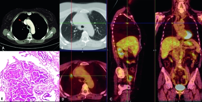
Chest x-rays of 270 patients with incidentally detected SPNs were retrospectively evaluated. Thirty patients with histologically proven SPNs were enrolled. On MDCT and positron emission tomography (PET)/CT images, two experts measured the density of nodules in all perfusion phases and the SUV. Net enhancement (NE) was calculated by subtracting peak pre-contrast density from peak post-contrast density. The Pearson test was used to correlate nodule NE, SUV, grading, histology and diameter.
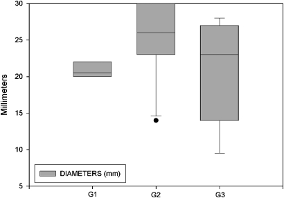
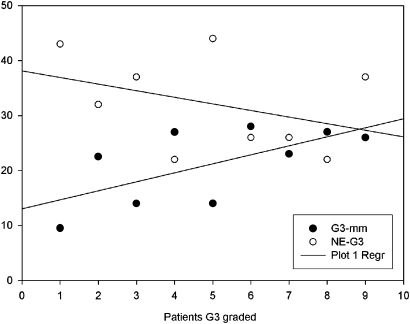
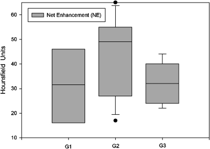
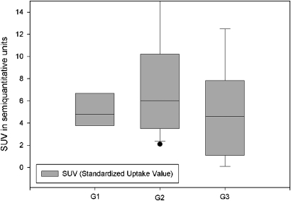
Of the 30 malignant SPNs, six were classified as G1 (median NE, 31.5 Hounsfield units (HU); median SUV, 4.8 units), 15 were classified as G2 (median NE, 49 HU; median SUV, 6 units), and nine were classified as G3 (median NE, 32 HU; median SUV, 4.5 units). A highly negative correlation was found in G3 SPNs between NE and the corresponding diameters (r=-0.834; p=0.00524). NE increased with the increase in diameter (r=0.982; p=0.284). SUV increased as the SPN diameter increased (r=0.789; p=0.421). NE and SUV were higher in G2 than G1 SPNs, and lower in G2 than G3 SPNs (r=0.97; p=0.137).
The significant correlation in dedifferentiated (G3) SPNs between NE and diameter (r=-0.834; p=0.00524) supports the theory that stroma and neoangiogenesis are fundamental in SPN growth. The highly negative correlation between NE and diameter demonstrates a net decrease in perfusion despite an increase in dimension. The multidisciplinary approach used herein may result in a more precise prognosis and consequently a better therapeutic outcome, particularly in patients with undifferentiated lung cancer.
评估孤立性肺结节(SPN)的组织学和分级是否与 30 例患者的动态多期多层 CT(MDCT)和[(18)F]氟脱氧葡萄糖标准化摄取值(SUV)结果相关。
回顾性评估了 270 例偶然发现的 SPN 患者的胸部 X 线片。纳入 30 例经组织学证实的 SPN 患者。在 MDCT 和正电子发射断层扫描(PET)/CT 图像上,两位专家测量了所有灌注期结节的密度和 SUV。通过从增强后峰值密度中减去增强前峰值密度来计算净增强(NE)。采用 Pearson 检验来关联结节的 NE、SUV、分级、组织学和直径。
30 例恶性 SPN 中,6 例为 G1(中位数 NE,31.5 亨氏单位(HU);中位数 SUV,4.8 单位),15 例为 G2(中位数 NE,49 HU;中位数 SUV,6 单位),9 例为 G3(中位数 NE,32 HU;中位数 SUV,4.5 单位)。在 G3 SPN 中,NE 与相应直径之间存在高度负相关(r=-0.834;p=0.00524)。随着 SPN 直径的增加,NE 也随之增加(r=0.982;p=0.284)。SUV 随着 SPN 直径的增加而增加(r=0.789;p=0.421)。与 G1 SPN 相比,G2 SPN 的 NE 和 SUV 较高,而 G3 SPN 的 NE 和 SUV 较低(r=0.97;p=0.137)。
分化不良(G3)SPN 中 NE 与直径之间的显著相关性(r=-0.834;p=0.00524)支持了基质和新生血管生成是 SPN 生长的基础理论。NE 与直径之间的高度负相关表明,尽管尺寸增加,但灌注净减少。本文采用的多学科方法可能会导致更精确的预后,从而带来更好的治疗效果,特别是在未分化肺癌患者中。