• 文献检索
  • 文档翻译
  • 深度研究
  • 学术资讯
  • Suppr Zotero 插件Zotero 插件
  • 邀请有礼
  • 套餐&价格
  • 历史记录
应用&插件
Suppr Zotero 插件Zotero 插件浏览器插件Mac 客户端Windows 客户端微信小程序
定价
高级版会员购买积分包购买API积分包
服务
文献检索文档翻译深度研究API 文档MCP 服务
关于我们
关于 Suppr公司介绍联系我们用户协议隐私条款
关注我们

Suppr 超能文献

核心技术专利:CN118964589B侵权必究
粤ICP备2023148730 号-1Suppr @ 2026

文献检索

告别复杂PubMed语法,用中文像聊天一样搜索,搜遍4000万医学文献。AI智能推荐,让科研检索更轻松。

立即免费搜索

文件翻译

保留排版,准确专业,支持PDF/Word/PPT等文件格式,支持 12+语言互译。

免费翻译文档

深度研究

AI帮你快速写综述,25分钟生成高质量综述,智能提取关键信息,辅助科研写作。

立即免费体验

视杯边缘中Wnt2b的表达需要色素上皮和非色素上皮之间的边界。

The expression of Wnt2b in the optic cup lip requires a border between the pigmented and nonpigmented epithelium.

作者信息

Kitamoto Junko, Hyer Jeanette

机构信息

Department of Ophthalmology and Neurosurgery, University of California, San Francisco, CA 94143, USA.

出版信息

Mol Vis. 2010 Dec 14;16:2701-17.

PMID:21179237
原文链接:https://pmc.ncbi.nlm.nih.gov/articles/PMC3002962/
Abstract

PURPOSE

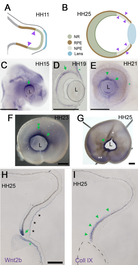
Wnt2b is normally expressed at the optic cup lip and is implicated in ciliary body induction. The lens has often been considered an organizer for the anterior eye, but recent studies demonstrate that the anterior cell fates are correctly specified in the absence of the lens. This study uses Wnt2b as a marker to reveal the mechanism behind the specification of the anterior domain of the optic cup.

METHODS

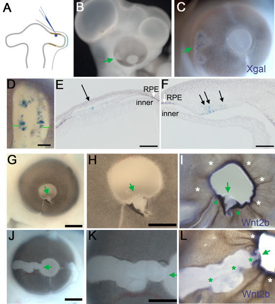
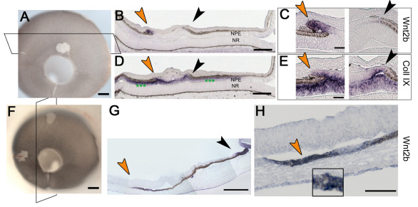
Developing chick embryos were used as a model system. Eyes were microsurgically manipulated to assess the role of the lens in the development of the anterior optic cup. Eyes were molecularly manipulated, using fibroblast growth factor expressing replication-incompetent retrovirus, introduced into the retinal pigmented epithelium (RPE) domain. Ectopic fibroblast growth factor transformed the RPE into nonpigmented epithelium (NPE; ciliary body). As the virus does not spread, discrete borders between RPE and NPE were experimentally created. Wnt2b expression was assessed after surgical and molecular manipulation.

RESULTS

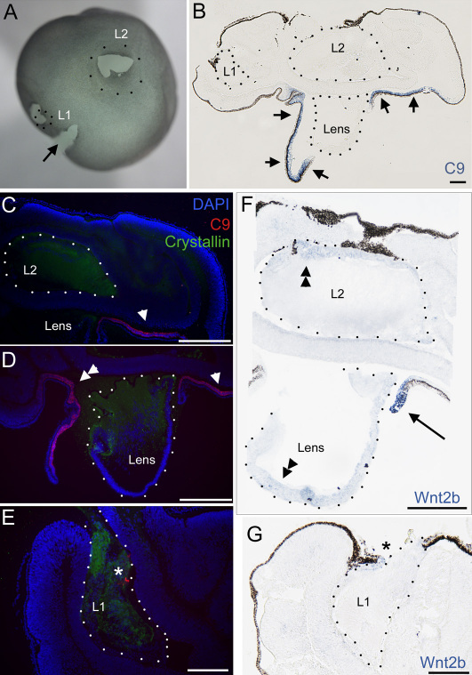
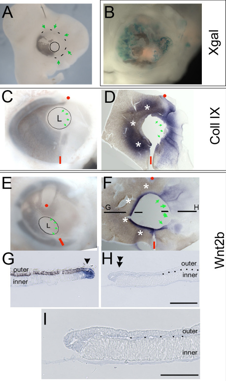
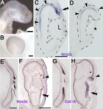
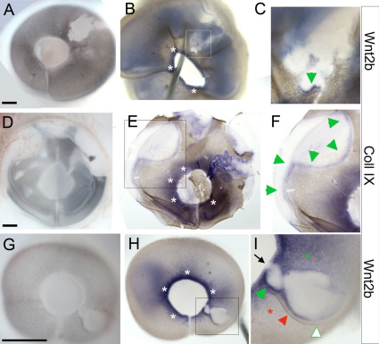
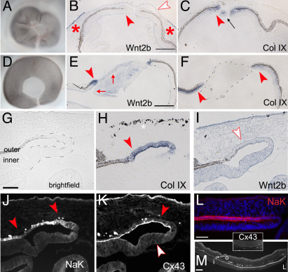
Contrary to expectations, we found that the lens is not able to induce Wnt2b expression in optic cup tissue: When the optic cup lip is experimentally misspecified such that it no longer contains the juxtaposition of pigmented and nonpigmented tissue, Wnt2b is not expressed. In addition, if the prelens ectoderm is removed from the optic vesicle before morphogenesis, the resulting lensless optic cup expresses Wnt2b even though it was not in contact with lens tissue. We also show that ectopic lenses do not induce Wnt2b in optic cup tissue. The ciliary body/anterior eye domain is specified at the border of RPE and the NPE of the ciliary body. During development, this border is normally found at the optic cup lip. We can manipulate tissue specification using retroviral-mediated gene transfer, and create ectopic borders between nonpigmented and pigmented tissue. At such borders, Wnt2b is ectopically expressed in the absence of lens contact. Finally, we describe a role for the lens in maintenance of Wnt2b expression and demonstrate support for this in two ways: First, we show that if the lens is removed from the formed optic cup, endogenous Wnt2b expression is specifically lost from the optic cup lip; and second, we show that while ectopic Wnt2b expression is initially found in the majority of ectopic borders, as eye development proceeds ectopic expression is maintained only in those borders that are close to the lens.

CONCLUSIONS

Taken together, the results provide support for a model in which the anterior optic cup domain, as described in part by Wnt2b expression, is specified through the elaboration of a border within the optic neuroepithelium rather than through interactions with the surrounding environment.

摘要
https://cdn.ncbi.nlm.nih.gov/pmc/blobs/93e1/3002962/4573b70b2ef3/mv-v16-2701-f8.jpg
https://cdn.ncbi.nlm.nih.gov/pmc/blobs/93e1/3002962/9307505c235c/mv-v16-2701-f1.jpg
https://cdn.ncbi.nlm.nih.gov/pmc/blobs/93e1/3002962/1e673d659eb9/mv-v16-2701-f2.jpg
https://cdn.ncbi.nlm.nih.gov/pmc/blobs/93e1/3002962/a3360605b37b/mv-v16-2701-f3.jpg
https://cdn.ncbi.nlm.nih.gov/pmc/blobs/93e1/3002962/517666658d1d/mv-v16-2701-f4.jpg
https://cdn.ncbi.nlm.nih.gov/pmc/blobs/93e1/3002962/bcabad01736b/mv-v16-2701-f5.jpg
https://cdn.ncbi.nlm.nih.gov/pmc/blobs/93e1/3002962/09bbf1cf8fb7/mv-v16-2701-f6.jpg
https://cdn.ncbi.nlm.nih.gov/pmc/blobs/93e1/3002962/c6f797e59122/mv-v16-2701-f7.jpg
https://cdn.ncbi.nlm.nih.gov/pmc/blobs/93e1/3002962/4573b70b2ef3/mv-v16-2701-f8.jpg

目的

Wnt2b通常在视杯边缘表达,并与睫状体诱导有关。晶状体常被认为是眼前部的组织者,但最近的研究表明,在没有晶状体的情况下,前部细胞命运也能正确指定。本研究使用Wnt2b作为标记物,以揭示视杯前部区域指定背后的机制。

方法

以发育中的鸡胚作为模型系统。通过显微手术对视进行操作,以评估晶状体在眼前部视杯发育中的作用。利用表达成纤维细胞生长因子的复制缺陷型逆转录病毒对视进行分子操作,并将其引入视网膜色素上皮(RPE)区域。异位的成纤维细胞生长因子将RPE转化为无色素上皮(NPE;睫状体)。由于病毒不会扩散,因此通过实验创建了RPE和NPE之间的离散边界。在手术和分子操作后评估Wnt2b的表达。

结果

与预期相反,我们发现晶状体无法诱导视杯组织中Wnt2b的表达:当通过实验错误指定视杯边缘,使其不再包含色素组织和无色素组织的并置时,Wnt2b不表达。此外,如果在形态发生之前从视泡中移除晶状体前外胚层,即使所得的无晶状体视杯未与晶状体组织接触,它也会表达Wnt2b。我们还表明,异位晶状体不会在视杯组织中诱导Wnt2b表达。睫状体/眼前部区域在睫状体的RPE和NPE的边界处指定。在发育过程中,这个边界通常位于视杯边缘。我们可以使用逆转录病毒介导的基因转移来操纵组织指定,并在无色素和色素组织之间创建异位边界。在这样的边界处,在没有晶状体接触的情况下,Wnt2b会异位表达。最后,我们描述了晶状体在维持Wnt2b表达中的作用,并通过两种方式证明了这一点:第一,我们表明,如果从形成的视杯中移除晶状体,视杯边缘会特异性地丧失内源性Wnt2b表达;第二,我们表明,虽然异位Wnt2b表达最初在大多数异位边界中发现,但随着眼睛发育的进行,异位表达仅在那些靠近晶状体的边界中维持。

结论

综上所述,这些结果支持了一个模型,即如部分由Wnt2b表达所描述的眼前部视杯区域,是通过视神经上皮内边界的形成来指定的,而不是通过与周围环境的相互作用。

相似文献

1
The expression of Wnt2b in the optic cup lip requires a border between the pigmented and nonpigmented epithelium.视杯边缘中Wnt2b的表达需要色素上皮和非色素上皮之间的边界。
Mol Vis. 2010 Dec 14;16:2701-17.
2
RPE specification in the chick is mediated by surface ectoderm-derived BMP and Wnt signalling.鸡的 RPE 特化由表面外胚层衍生的 BMP 和 Wnt 信号介导。
Development. 2013 Dec;140(24):4959-69. doi: 10.1242/dev.096990. Epub 2013 Nov 13.
3
FGF-mediated induction of ciliary body tissue in the chick eye.成纤维细胞生长因子介导的鸡眼中睫状体组织的诱导
Dev Biol. 2007 Apr 1;304(1):272-85. doi: 10.1016/j.ydbio.2006.12.033. Epub 2006 Dec 21.
4
Optic cup morphogenesis requires pre-lens ectoderm but not lens differentiation.视杯形态发生需要晶状体前外胚层,但不需要晶状体分化。
Dev Biol. 2003 Jul 15;259(2):351-63. doi: 10.1016/s0012-1606(03)00205-7.
5
Bmp4 from the optic vesicle specifies murine retina formation.来自视泡的骨形态发生蛋白4决定小鼠视网膜的形成。
Dev Biol. 2015 Jun 1;402(1):119-26. doi: 10.1016/j.ydbio.2015.03.006. Epub 2015 Mar 16.
6
Eye morphogenesis and patterning of the optic vesicle.眼球形态发生和视泡的模式形成。
Curr Top Dev Biol. 2010;93:61-84. doi: 10.1016/B978-0-12-385044-7.00003-5.
7
Retinal and anterior eye compartments derive from a common progenitor pool in the avian optic cup.视网膜和眼前节源自禽类视杯中的一个共同祖细胞池。
Mol Vis. 2011;17:3347-63. Epub 2011 Dec 20.
8
Otx2 is involved in the regional specification of the developing retinal pigment epithelium by preventing the expression of sox2 and fgf8, factors that induce neural retina differentiation.Otx2 通过防止 Sox2 和 Fgf8 的表达来参与发育中的视网膜色素上皮的区域特化,这两种因子诱导神经视网膜分化。
PLoS One. 2012;7(11):e48879. doi: 10.1371/journal.pone.0048879. Epub 2012 Nov 8.
9
Coming into focus: the role of extracellular matrix in vertebrate optic cup morphogenesis.逐渐明晰:细胞外基质在脊椎动物视杯形态发生中的作用
Dev Dyn. 2014 Oct;243(10):1242-8. doi: 10.1002/dvdy.24162. Epub 2014 Jul 19.
10
Bone morphogenetic proteins specify the retinal pigment epithelium in the chick embryo.骨形态发生蛋白决定了鸡胚中的视网膜色素上皮。
Development. 2007 Oct;134(19):3483-93. doi: 10.1242/dev.02884. Epub 2007 Aug 29.

引用本文的文献

1
The development and the genetic diseases of the ciliary body.睫状体的发育与遗传性疾病。
Cell Insight. 2024 Mar 15;3(3):100162. doi: 10.1016/j.cellin.2024.100162. eCollection 2024 Jun.
2
BMP-induced reprogramming of the neural retina into retinal pigment epithelium requires Wnt signalling.骨形态发生蛋白诱导神经视网膜重编程为视网膜色素上皮需要Wnt信号通路。
Biol Open. 2017 Jul 15;6(7):979-992. doi: 10.1242/bio.018739.
3
Establishment of the neurogenic boundary of the mouse retina requires cooperation of SOX2 and WNT signaling.

本文引用的文献

1
Distribution of Pax6 protein during eye development suggests discrete roles in proliferative and differentiated visual cells.眼发育过程中Pax6蛋白的分布表明其在增殖性和分化性视觉细胞中具有不同作用。
Dev Genes Evol. 1997 Jan;206(6):363-369. doi: 10.1007/s004270050065.
2
Spatial and temporal regulation of Wnt/beta-catenin signaling is essential for development of the retinal pigment epithelium.Wnt/β-连环蛋白信号通路的时空调控对于视网膜色素上皮的发育至关重要。
Dev Biol. 2009 Oct 1;334(1):31-45. doi: 10.1016/j.ydbio.2009.07.002. Epub 2009 Jul 9.
3
Beta-catenin controls differentiation of the retinal pigment epithelium in the mouse optic cup by regulating Mitf and Otx2 expression.
小鼠视网膜神经源性边界的建立需要SOX2和WNT信号的协同作用。
Neural Dev. 2014 Dec 9;9:27. doi: 10.1186/1749-8104-9-27.
4
β-Catenin inactivation is a pre-requisite for chick retina regeneration.β-连环蛋白失活是鸡视网膜再生的一个先决条件。
PLoS One. 2014 Jul 8;9(7):e101748. doi: 10.1371/journal.pone.0101748. eCollection 2014.
5
Identification of Wnt/β-catenin modulated genes in the developing retina.发育中的视网膜中Wnt/β-连环蛋白调控基因的鉴定。
Mol Vis. 2012;18:645-56. Epub 2012 Mar 16.
6
Retinal and anterior eye compartments derive from a common progenitor pool in the avian optic cup.视网膜和眼前节源自禽类视杯中的一个共同祖细胞池。
Mol Vis. 2011;17:3347-63. Epub 2011 Dec 20.
β-连环蛋白通过调节小眼畸形相关转录因子(Mitf)和正交同源盒2(Otx2)的表达来控制小鼠视杯视网膜色素上皮的分化。
Development. 2009 Aug;136(15):2505-10. doi: 10.1242/dev.032136. Epub 2009 Jun 24.
4
Hairy1 acts as a node downstream of Wnt signaling to maintain retinal stem cell-like progenitor cells in the chick ciliary marginal zone.Hairy1作为Wnt信号下游的一个节点,维持鸡睫状边缘区的视网膜干细胞样祖细胞。
Development. 2009 Jun;136(11):1823-33. doi: 10.1242/dev.029272. Epub 2009 Apr 22.
5
Depression of intraocular pressure following inactivation of connexin43 in the nonpigmented epithelium of the ciliary body.睫状体非色素上皮中连接蛋白43失活后眼压降低。
Invest Ophthalmol Vis Sci. 2009 May;50(5):2185-93. doi: 10.1167/iovs.08-2962. Epub 2009 Jan 24.
6
Wnt signaling in eye organogenesis.Wnt 信号在眼器官发生中的作用。
Organogenesis. 2008 Apr;4(2):60-7. doi: 10.4161/org.4.2.5850.
7
Fgf8a induces neural crest indirectly through the activation of Wnt8 in the paraxial mesoderm.Fgf8a通过激活近轴中胚层中的Wnt8间接诱导神经嵴。
Development. 2008 Dec;135(23):3903-10. doi: 10.1242/dev.026229.
8
Characterization of a transient TCF/LEF-responsive progenitor population in the embryonic mouse retina.胚胎小鼠视网膜中瞬时TCF/LEF反应性祖细胞群体的特征分析。
Invest Ophthalmol Vis Sci. 2009 Jan;50(1):432-40. doi: 10.1167/iovs.08-2270. Epub 2008 Jul 3.
9
Perinatal ablation of the mouse lens causes multiple anterior chamber defects.围产期对小鼠晶状体进行消融会导致多个前房缺陷。
Mol Vis. 2007 Dec 18;13:2289-300.
10
Activin signaling limits the competence for retinal regeneration from the pigmented epithelium.激活素信号传导限制了色素上皮细胞进行视网膜再生的能力。
Mech Dev. 2008 Jan-Feb;125(1-2):106-16. doi: 10.1016/j.mod.2007.10.001. Epub 2007 Oct 10.