Breast Tumor Center, Sun Yat-Sen Memorial Hospital, Sun Yat-Sen University, Guangzhou, People's Republic of China.
PLoS One. 2010 Dec 20;5(12):e15630. doi: 10.1371/journal.pone.0015630.
Evidence is lacking whether the number of breast tumor-initiating cells (BT-ICs) directly correlates with the sensitivity of breast tumors to chemotherapy. Here, we evaluated the association between proportion of BT-ICs and chemoresistance of the tumors.
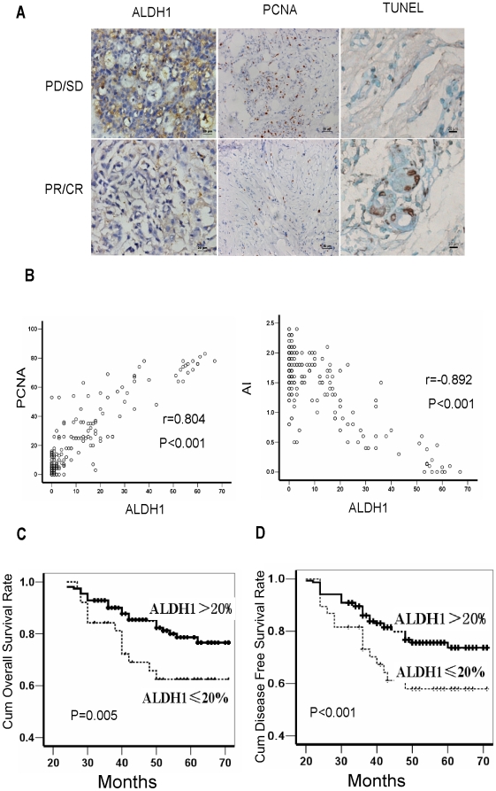
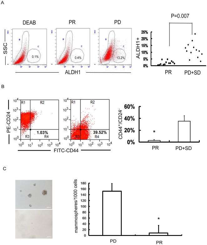
Immunohistochemical staining(IHC) was used to examine the expression of aldehyde dehydrogenase 1 (ALDH1) and proliferating cell nuclear antigen, and TUNEL was used to detect the apoptosis index. The significance of various variables in patient survival was analyzed using a Cox proportional hazards model. The percentage of BT-ICs in breast cancer cell lines and primary breast tumors was determined by ALDH1 enzymatic assay, CD44(+)/CD24(-) phenotype and mammosphere formation assay.
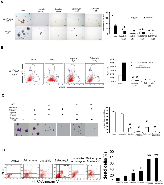
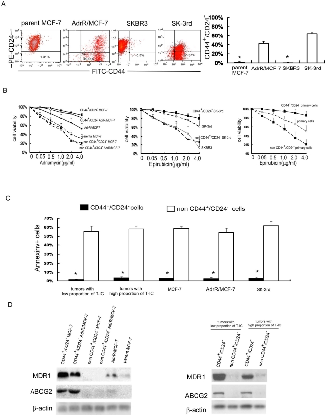
ALDH1 expression determined by IHC in primary breast cancers was associated with poor clinical response to neoadjuvant chemotherapy and reduced survival in breast cancer patients. Breast tumors that contained higher proportion of BT-ICs with CD44(+)/CD24(-) phenotype, ALDH1 enzymatic activity and sphere forming capacity were more resistant to neoadjuvant chemotherapy. Chemoresistant cell lines AdrR/MCF-7 and SK-3rd, had increased number of cells with sphere forming capacity, CD44(+)/CD24(-) phenotype and side-population. Regardless the proportion of T-ICs, FACS-sorted CD44(+)/CD24(-) cells that derived from primary tumors or breast cancer lines were about 10-60 fold more resistant to chemotherapy relative to the non- CD44(+)/CD24(-) cells and their parental cells. Furthermore, our data demonstrated that MDR1 (multidrug resistance 1) and ABCG2 (ATP-binding cassette sub-family G member 2) were upregulated in CD44(+)/CD24(-) cells. Treatment with lapatinib or salinomycin reduced the proportion of BT-ICs by nearly 50 fold, and thus enhanced the sensitivity of breast cancer cells to chemotherapy by around 30 fold.
These data suggest that the proportion of BT-ICs is associated with chemotherapeutic resistance of breast cancer. It highlights the importance of targeting T-ICs, rather than eliminating the bulk of rapidly dividing and terminally differentiated cells, in novel anti-cancer strategies.
目前尚缺乏证据表明乳腺癌肿瘤起始细胞(BT-ICs)的数量是否与肿瘤对化疗的敏感性直接相关。在这里,我们评估了 BT-ICs 比例与肿瘤化疗耐药性之间的关系。
采用免疫组化染色(IHC)检测醛脱氢酶 1(ALDH1)和增殖细胞核抗原的表达,采用 TUNEL 检测细胞凋亡指数。采用 Cox 比例风险模型分析患者生存中各种变量的意义。采用 ALDH1 酶法、CD44(+)/CD24(-)表型和乳腺球体形成试验确定乳腺癌细胞系和原发性乳腺癌中 BT-ICs 的百分比。
原发性乳腺癌中 IHC 测定的 ALDH1 表达与新辅助化疗的临床反应不良和乳腺癌患者生存时间缩短有关。含有较高 CD44(+)/CD24(-)表型、ALDH1 酶活性和球体形成能力的 BT-ICs 比例较高的乳腺肿瘤对新辅助化疗的耐药性更强。耐药细胞系 AdrR/MCF-7 和 SK-3rd 具有更多具有球体形成能力、CD44(+)/CD24(-)表型和侧群的细胞。无论 T-ICs 的比例如何,FACS 分选的源自原发性肿瘤或乳腺癌系的 CD44(+)/CD24(-)细胞对化疗的耐药性约为非 CD44(+)/CD24(-)细胞及其亲本细胞的 10-60 倍。此外,我们的数据表明 MDR1(多药耐药 1)和 ABCG2(ATP 结合盒亚家族 G 成员 2)在 CD44(+)/CD24(-)细胞中上调。用拉帕替尼或沙利霉素处理可使 BT-ICs 的比例降低近 50 倍,从而使乳腺癌细胞对化疗的敏感性提高约 30 倍。
这些数据表明 BT-ICs 的比例与乳腺癌的化疗耐药性有关。这强调了在新的抗癌策略中靶向 T-ICs 而不是消除快速分裂和终末分化细胞的重要性。