Jung Da Eun, Lee Joon-Soo
Department of Pediatrics, Ajou University School of Medicine, Suwon, Korea.
Korean J Pediatr. 2010 Aug;53(8):779-85. doi: 10.3345/kjp.2010.53.8.779. Epub 2010 Aug 31.
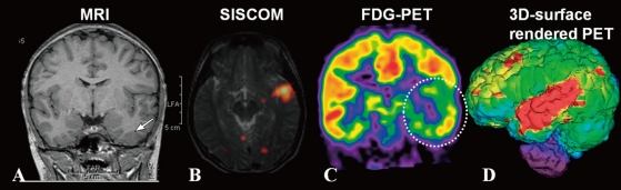
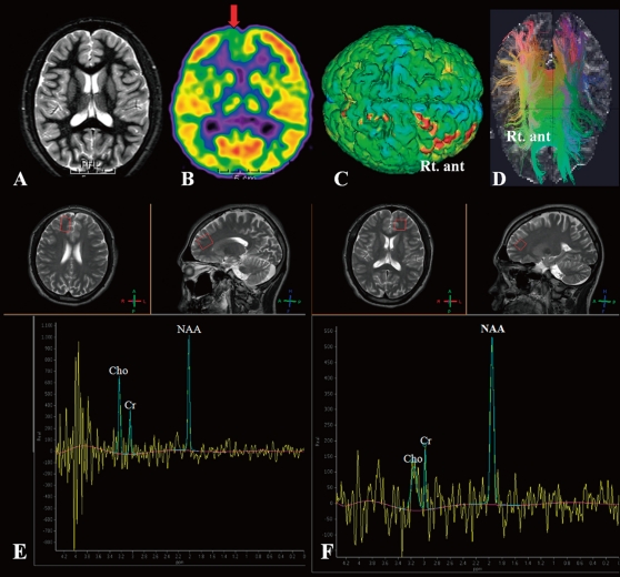
In pre-surgical evaluation of pediatric epilepsy, the combined use of multiple imaging modalities for precise localization of the epileptogenic focus is a worthwhile endeavor. Advanced neuroimaging by high field Magnetic resonance imaging (MRI), diffusion tensor images, and MR spectroscopy have the potential to identify subtle lesions. (18)F-FDG positron emission tomography and single photon emission tomography provide visualization of metabolic alterations of the brain in the ictal and interictal states. These techniques may have localizing value for patients which exhibit normal MRI scans. Functional MRI is helpful for non-invasively identifying areas of eloquent cortex. These advances are improving our ability to noninvasively detect epileptogenic foci which have gone undetected in the past and whose accurate localization is crucial for a favorable outcome following surgical resection.
在小儿癫痫的术前评估中,联合使用多种成像方式以精确确定致痫灶的位置是一项值得努力的工作。高场磁共振成像(MRI)、弥散张量成像和磁共振波谱等先进的神经成像技术有潜力识别细微病变。(18)F-FDG正电子发射断层扫描和单光子发射断层扫描可显示发作期和发作间期大脑的代谢改变。对于MRI扫描正常的患者,这些技术可能具有定位价值。功能MRI有助于无创识别明确的皮质区域。这些进展正在提高我们无创检测过去未被发现的致痫灶的能力,而其精确定位对于手术切除后的良好预后至关重要。