Department of Pathology and Molecular Pathology.
J Pain Res. 2010 Nov 8;3:213-21. doi: 10.2147/JPR.S14209.
The objectives of this study were to establish and characterize a novel animal model of metastatic prostate cancer-induced bone pain.
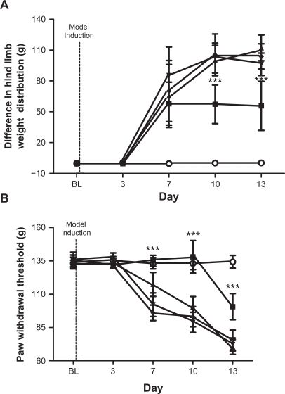
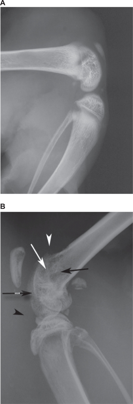
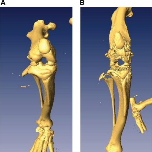
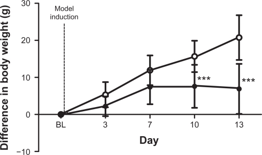
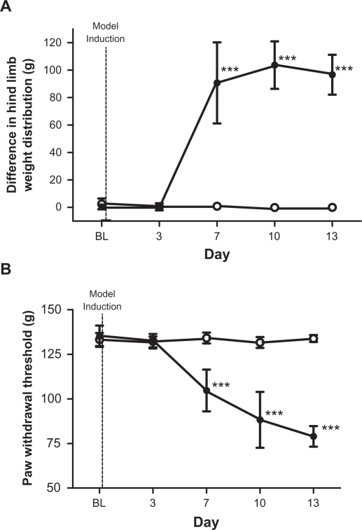
Copenhagen rats were injected with 10(6) MATLyLu (MLL) prostate cancer cells or phosphate-buffered saline by per cutaneous intra femoral injections into the right hind leg distal epiphysis. Over 13 days, rats progressively developed a tumor within the distal femoral epiphysis. On days 3, 7, 10, and 13 post injection, rats were subjected to the incapacitance and Randall-Selitto behavioral tests as they are believed to be indirect reflections of tumor induced pain. Ipsilateral hind limbs were subjected to X-ray and computed tomography (CT) scans and histological sections were stained with hematoxylin and eosin (H&E).
Intra femoral injections of MLL cells resulted in the progressive development of a tumor leading to bone destruction and nociceptive behaviors. Tumor development resulted in the redistribution of weight to the contralateral hind leg and significantly reduced the paw withdrawal threshold of the ipsilateral hind paw as observed via the incapacitance and Randall-Selitto tests, respectively. X-ray and computed tomography scans along with H&E stains indicated tumor-associated structural damage to the distal femur. This model was challenged with administration of meloxicam. Compared with vehicle-injected controls, the meloxicam-treated rats displayed smaller nociceptive responses as observed with the incapacitance and Randall-Selitto tests, suggesting that meloxicam was effective in reducing the pain-related symptoms displayed by model animals and that the model behaved in a predictable way to cyclooxygenase-2 treatment.
This model is unique from other bone cancer models in that it is a comprehensive model utilizing a competent immune system with a syngeneic tumor. The model establishes a tool that will be useful to investigate mechanisms of cancer pain that are induced by cancer cells.
本研究的目的是建立并描述一种新型转移性前列腺癌诱导骨痛动物模型。
通过经皮股骨内向右侧后腿远侧骺内注射,将 10(6)MATLyLu(MLL)前列腺癌细胞或磷酸盐缓冲盐水注入哥本哈根大鼠。在 13 天内,大鼠的远侧股骨骺内逐渐形成肿瘤。在注射后第 3、7、10 和 13 天,大鼠进行了无法容忍和兰德尔-塞利托行为测试,因为它们被认为是肿瘤诱导疼痛的间接反映。同侧后肢进行 X 射线和计算机断层扫描(CT)扫描,并用苏木精和伊红(H&E)染色对组织切片进行染色。
MLL 细胞的股骨内向注射导致肿瘤的进行性发展,导致骨破坏和伤害性行为。肿瘤的发展导致体重重新分布到对侧后腿,并显著降低了同侧后腿的足撤回阈值,分别通过无法容忍和兰德尔-塞利托测试观察到。X 射线和计算机断层扫描以及 H&E 染色表明肿瘤相关的对远侧股骨的结构损伤。该模型接受了美洛昔康的给药。与 vehicle-injected 对照组相比,美洛昔康治疗组大鼠在无法容忍和兰德尔-塞利托测试中表现出较小的伤害性反应,这表明美洛昔康在减轻模型动物的疼痛相关症状方面有效,并且该模型对环氧化酶-2 治疗表现出可预测的行为。
与其他骨癌模型相比,该模型具有独特性,因为它是一种利用具有同源肿瘤的功能免疫系统的综合模型。该模型建立了一种工具,将有助于研究由癌细胞引起的癌症疼痛的机制。