Molecular Genetics Dept., Weizmann Inst., Israel.
Int J Biol Sci. 2010 Dec 26;7(1):1-8. doi: 10.7150/ijbs.7.1.
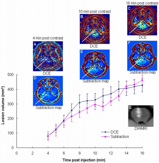
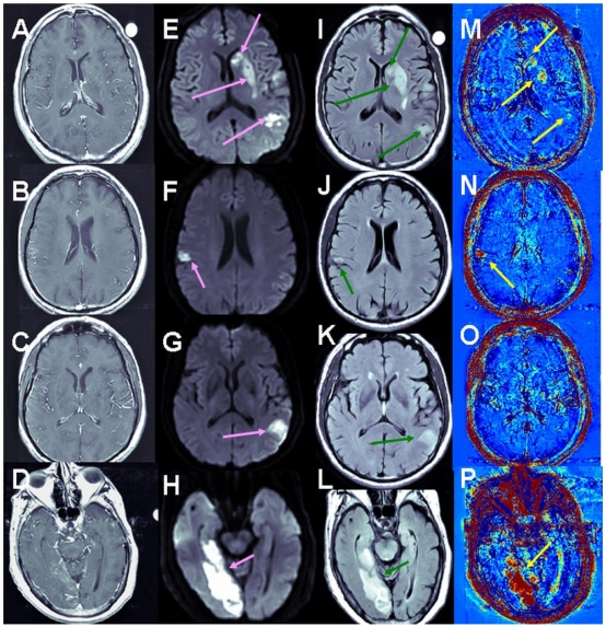
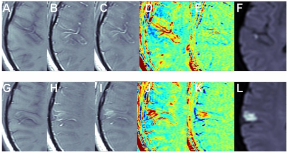
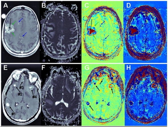
The development of imaging methodologies for detecting blood-brain-barrier (BBB) disruption may help predict stroke patient's propensity to develop hemorrhagic complications following reperfusion. We have developed a delayed contrast extravasation MRI-based methodology enabling real-time depiction of subtle BBB abnormalities in humans with high sensitivity to BBB disruption and high spatial resolution. The increased sensitivity to subtle BBB disruption is obtained by acquiring T1-weighted MRI at relatively long delays (~15 minutes) after contrast injection and subtracting from them images acquired immediately after contrast administration. In addition, the relatively long delays allow for acquisition of high resolution images resulting in high resolution BBB disruption maps. The sensitivity is further increased by image preprocessing with corrections for intensity variations and with whole body (rigid+elastic) registration. Since only two separate time points are required, the time between the two acquisitions can be used for acquiring routine clinical data, keeping the total imaging time to a minimum. A proof of concept study was performed in 34 patients with ischemic stroke and 2 patients with brain metastases undergoing high resolution T1-weighted MRI acquired at 3 time points after contrast injection. The MR images were pre-processed and subtracted to produce BBB disruption maps. BBB maps of patients with brain metastases and ischemic stroke presented different patterns of BBB opening. The significant advantage of the long extravasation time was demonstrated by a dynamic-contrast-enhancement study performed continuously for 18 min. The high sensitivity of our methodology enabled depiction of clear BBB disruption in 27% of the stroke patients who did not have abnormalities on conventional contrast-enhanced MRI. In 36% of the patients, who had abnormalities detectable by conventional MRI, the BBB disruption volumes were significantly larger in the maps than in conventional MRI. These results demonstrate the advantages of delayed contrast extravasation in increasing the sensitivity to subtle BBB disruption in ischemic stroke patients. The calculated disruption maps provide clear depiction of significant volumes of BBB disruption unattainable by conventional contrast-enhanced MRI.
成像方法的发展用于检测血脑屏障(BBB)破坏可能有助于预测中风患者在再灌注后发生出血性并发症的倾向。我们开发了一种基于对比延迟外渗的 MRI 方法,能够实时描绘人类 BBB 细微异常,对 BBB 破坏具有高灵敏度和高空间分辨率。通过在对比注射后相对较长的延迟(约 15 分钟)采集 T1 加权 MRI,并从它们中减去立即在对比给药后采集的图像,可以获得对细微 BBB 破坏的更高灵敏度。此外,相对较长的延迟允许采集高分辨率图像,从而产生高分辨率的 BBB 破坏图。通过对图像进行预处理,包括强度变化的校正和全身(刚性+弹性)配准,可以进一步提高灵敏度。由于只需要两个单独的时间点,因此两次采集之间的时间可以用于获取常规临床数据,使总成像时间最小化。在 34 例缺血性中风患者和 2 例脑转移患者中进行了概念验证研究,这些患者在对比注射后 3 个时间点进行了高分辨率 T1 加权 MRI 采集。对 MR 图像进行预处理和相减,以产生 BBB 破坏图。脑转移和缺血性中风患者的 BBB 图呈现出不同的 BBB 开放模式。通过连续进行 18 分钟的动态对比增强研究,证明了长外渗时间的显著优势。我们的方法具有很高的灵敏度,能够在 27%的没有常规对比增强 MRI 异常的中风患者中清晰地显示 BBB 破坏。在 36%的患者中,常规 MRI 可检测到异常,BBB 破坏体积在图中明显大于常规 MRI。这些结果表明,延迟对比外渗在增加对缺血性中风患者细微 BBB 破坏的灵敏度方面具有优势。计算出的破坏图清晰地描绘了常规对比增强 MRI 无法获得的大量 BBB 破坏。