Department of Pharmaceutical Sciences, University of Arkansas for Medical Sciences, 4301 W. Markham St., #522-3, Little Rock, AR 72205-7199, USA.
Brain Res. 2011 Mar 10;1378:54-65. doi: 10.1016/j.brainres.2011.01.028. Epub 2011 Jan 15.
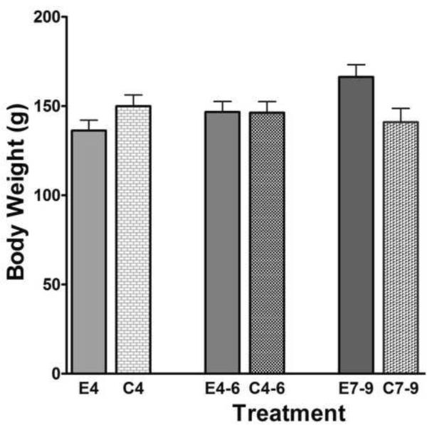
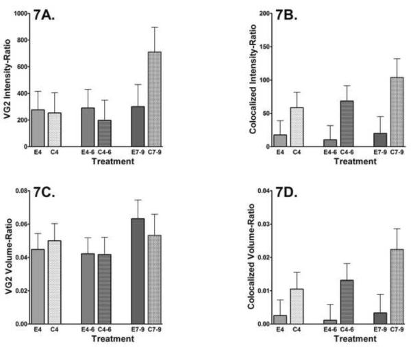
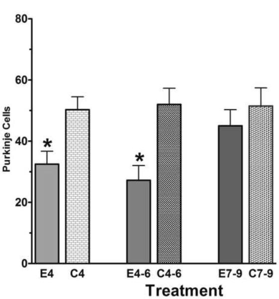
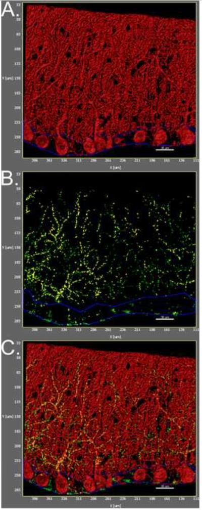
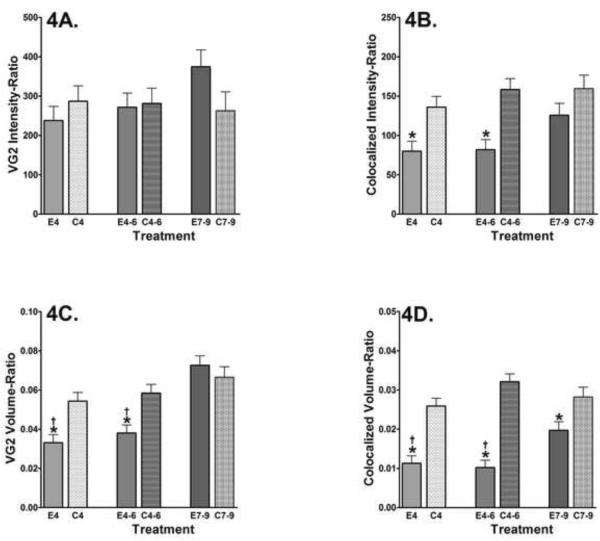
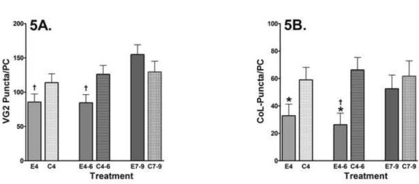
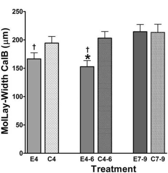
Developmental ethanol exposure in rats during postnatal days (PN) 4-6 is known to cause significant loss of the cerebellar Purkinje cells. It is not known what happens to the surviving neurons as they continue to develop. This study was designed to quantify the interactions between the olivary climbing fibers and the Purkinje cells when the cerebellar circuits have matured. Rat pups were treated with a daily dose of ethanol (4.5g/kg body weight) delivered by intragastric intubation on PN4, PN4-6, or PN7-9. The interactions between the climbing fibers and the Purkinje cells were examined on PN40 using confocal microscopy. Mid-vermal cerebellar sections were stained with antibodies to calbindin-D28k (to visualize Purkinje cells) and vesicular glutamate transporter 2 (VGluT2, to visualize climbing fibers). Confocal z-stack images were obtained from Lobule 1 and analyzed with Imaris software to quantify the staining of the two antibodies. The VGluT2 immunostaining was significantly reduced and this was associated with alterations in the synaptic integrity, and synaptic number per Purkinje cell with only a single exposure on PN4 enough to cause the alterations. Previously, we demonstrated similar deficits in climbing fiber innervation when analyzed on PN14 (Pierce, Hayar, Williams, and Light, 2010). The present study confirms that these alterations are sustained and further identifies the decreased synaptic density as well as alterations to the general morphology of the molecular layer of the cerebellar cortex that are the result of the binge ethanol exposure.
发育期(新生后第 4-6 天)给予大鼠乙醇暴露会导致明显的小脑浦肯野细胞丢失。目前尚不清楚存活的神经元在继续发育时会发生什么。本研究旨在定量分析小脑回路成熟后橄榄核攀援纤维与浦肯野细胞之间的相互作用。新生大鼠在新生后第 4 天、第 4-6 天或第 7-9 天通过胃内插管给予每日剂量的乙醇(4.5g/kg 体重)。在新生后第 40 天,使用共聚焦显微镜检查攀援纤维与浦肯野细胞之间的相互作用。用钙结合蛋白-D28k(用于可视化浦肯野细胞)和囊泡谷氨酸转运体 2(VGluT2,用于可视化攀援纤维)抗体对小脑中间叶切片进行染色。从 1 叶获得共聚焦 z 堆叠图像,并使用 Imaris 软件对其进行分析,以定量两种抗体的染色。VGluT2 免疫染色显著减少,这与突触完整性改变以及每个浦肯野细胞的突触数量减少有关,仅在新生后第 4 天单次暴露就足以引起这些改变。之前,我们在分析新生后第 14 天的攀援纤维支配时也发现了类似的缺陷(Pierce、Hayar、Williams 和 Light,2010)。本研究证实这些改变是持续的,并进一步确定了突触密度降低以及小脑皮质分子层的一般形态改变,这些都是 binge 乙醇暴露的结果。