Ward Heather H, Romero Elsa, Welford Angela, Pickett Gavin, Bacallao Robert, Gattone Vincent H, Ness Scott A, Wandinger-Ness Angela, Roitbak Tamara
Department of Pathology, University of New Mexico Health Sciences Center, Albuquerque, NM 87131, USA.
Biochim Biophys Acta. 2011 Oct;1812(10):1344-57. doi: 10.1016/j.bbadis.2011.01.010. Epub 2011 Jan 19.
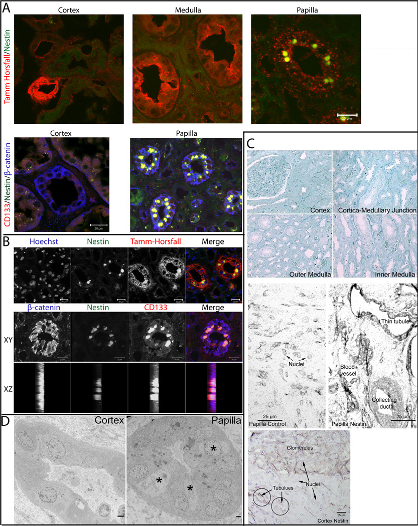
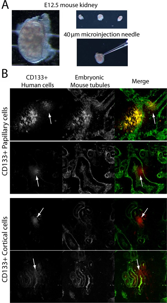
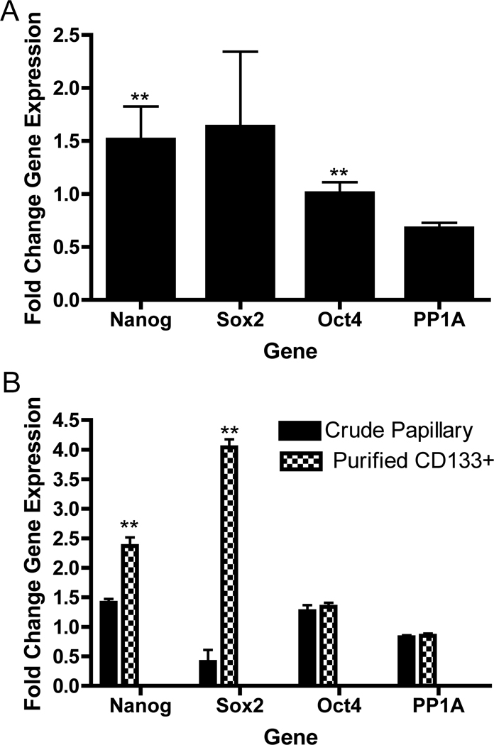
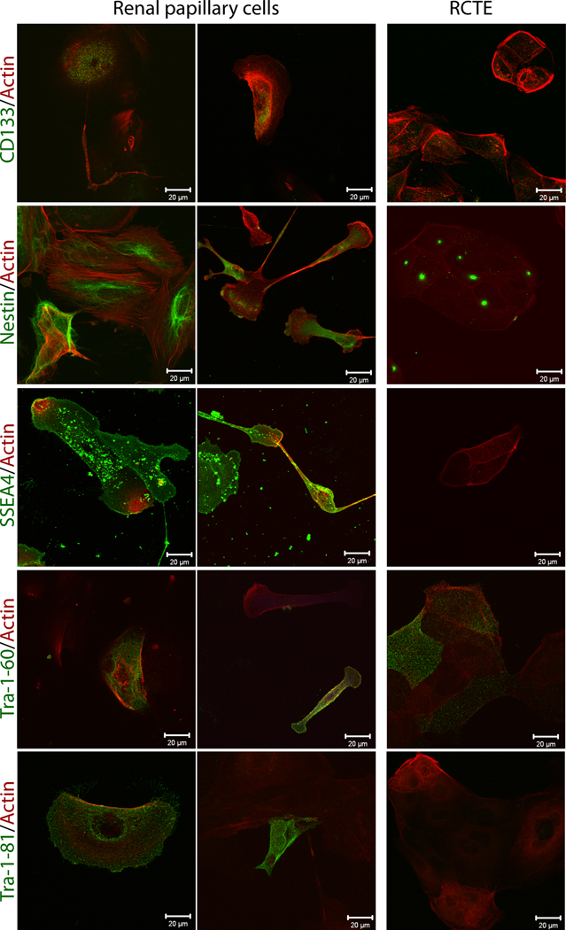
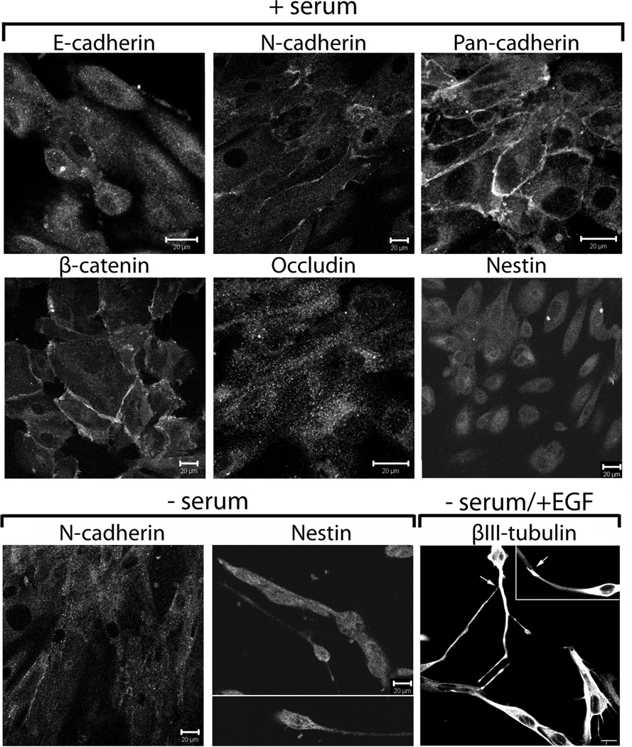
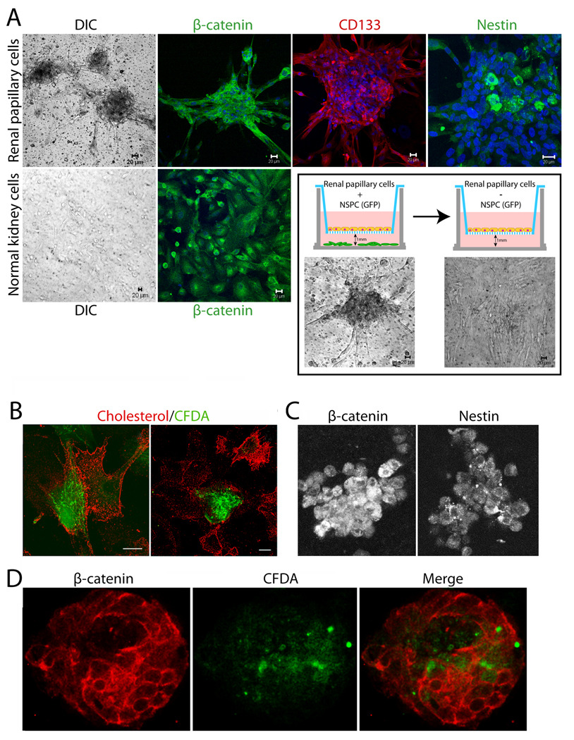
Approximately 60,000 patients in the United States are waiting for a kidney transplant due to genetic, immunologic and environmentally caused kidney failure. Adult human renal stem cells could offer opportunities for autologous transplant and repair of damaged organs. Current data suggest that there are multiple progenitor types in the kidney with distinct localizations. In the present study, we characterize cells derived from human kidney papilla and show their capacity for tubulogenesis. In situ, nestin(+) and CD133/1(+) cells were found extensively intercalated between tubular epithelia in the loops of Henle of renal papilla, but not of the cortex. Populations of primary cells from the renal cortex and renal papilla were isolated by enzymatic digestion from human kidneys unsuited for transplant and immuno-enriched for CD133/1(+) cells. Isolated CD133/1(+) papillary cells were positive for nestin, as well as several human embryonic stem cell markers (SSEA4, Nanog, SOX2, and OCT4/POU5F1) and could be triggered to adopt tubular epithelial and neuronal-like phenotypes. Isolated papillary cells exhibited morphologic plasticity upon modulation of culture conditions and inhibition of asymmetric cell division. Labeled papillary cells readily associated with cortical tubular epithelia in co-culture and 3-dimensional collagen gel cultures. Heterologous organ culture demonstrated that CD133/1(+) progenitors from the papilla and cortex became integrated into developing kidney tubules. Tubular epithelia did not participate in tubulogenesis. Human renal papilla harbor cells with the hallmarks of adult kidney stem/progenitor cells that can be amplified and phenotypically modulated in culture while retaining the capacity to form new kidney tubules. This article is part of a Special Issue entitled: Polycystic Kidney Disease.
在美国,约有6万名患者因遗传、免疫和环境因素导致肾衰竭而等待肾移植。成人肾干细胞可为自体移植和受损器官修复提供机会。目前的数据表明,肾脏中存在多种定位不同的祖细胞类型。在本研究中,我们对源自人肾乳头的细胞进行了表征,并展示了它们的成管能力。在原位,巢蛋白(+)和CD133/1(+)细胞广泛插在肾乳头而非皮质的髓袢肾小管上皮之间。通过酶消化从不适合移植的人肾脏中分离出肾皮质和肾乳头的原代细胞群体,并对CD133/1(+)细胞进行免疫富集。分离出的CD133/1(+)乳头细胞对巢蛋白呈阳性,同时对几种人类胚胎干细胞标志物(SSEA4、Nanog、SOX2和OCT4/POU5F1)也呈阳性,并且可以被触发呈现肾小管上皮和神经元样表型。分离出的乳头细胞在培养条件调节和不对称细胞分裂抑制后表现出形态可塑性。在共培养和三维胶原凝胶培养中,标记的乳头细胞很容易与皮质肾小管上皮结合。异源器官培养表明,来自乳头和皮质的CD133/1(+)祖细胞可整合到发育中的肾小管中。肾小管上皮不参与成管过程。人肾乳头含有具有成人肾干细胞/祖细胞特征的细胞,这些细胞在培养中可以扩增并进行表型调节,同时保留形成新肾小管的能力。本文是名为:多囊肾病的特刊的一部分。