Department of Biology, University of York, York YO10 5DD, UK.
Br J Cancer. 2011 Feb 15;104(4):673-84. doi: 10.1038/sj.bjc.6606077. Epub 2011 Jan 25.
Expression of protein kinase C alpha (PKCα) is elevated in prostate cancer (PCa); thus, we have studied whether the development of tumourigenesis in prostate epithelial cell lines modifies the normal pattern of choline (Cho) metabolite release on PKC activation.
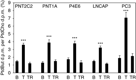
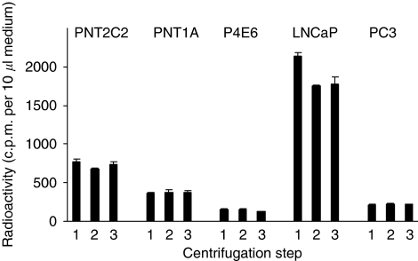
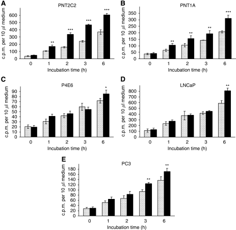
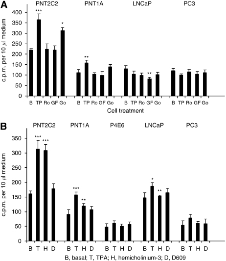
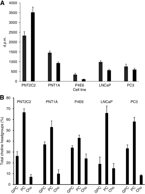
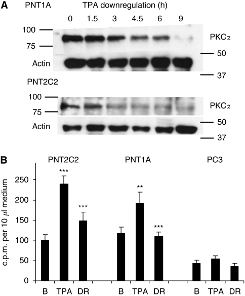
Normal and tumourigenic human prostate epithelial cell lines were incubated with [(3)H]-Cho to label choline phospholipids. Protein kinase C was activated with phorbol ester and blocked with inhibitors. Choline metabolites were resolved by ion-exchange chromatography. Phospholipase D (PLD) activity was measured by transphosphatidylation. Protein expression was detected by western blotting and/or RT-PCR. Choline uptake was measured on cells in monolayers over 60 min.
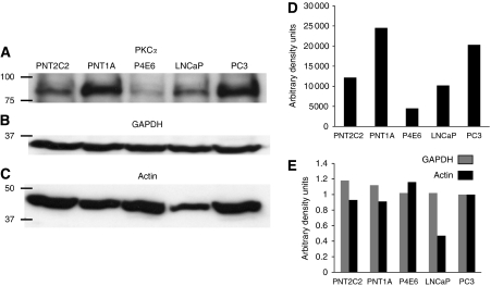
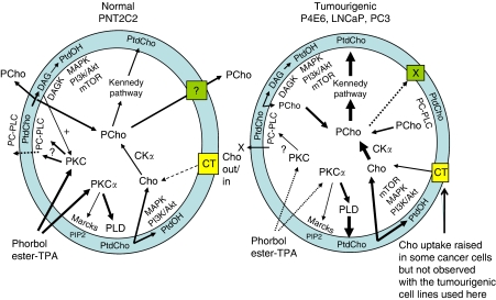
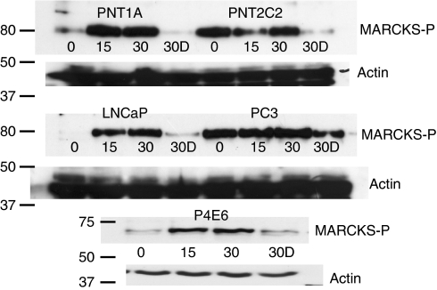
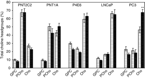
Normal prostate epithelial cell lines principally released phosphocholine (PCho) in contrast to tumourigenic lines, which released Cho. In addition, only with normal cell lines did PKC activation stimulate Cho metabolite release. Protein kinase C alpha expression varied between normal and tumourigenic cell lines but all showed a PKCα link to myristoylated alanine-rich C kinase substrate (MARCKS) protein. The five cell lines differed in Cho uptake levels, with normal PNT2C2 line cells showing highest uptake over 60 min incubation. Normal and tumourigenic cell lines expressed mRNA for PLD1 and PLD2, and showed similar levels of basal and PKC-activated PLD activity.
The transition to tumourigenesis in prostate epithelial cell lines results in major changes to Cho metabolite release into the medium and PKC signalling to phosphatidylcholine turnover. The changes, which reflect the metabolic and proliferative needs of tumourigenic cells compared with untransformed cells, could be significant for both diagnosis and treatment.
蛋白激酶 C 阿尔法(PKCα)的表达在前列腺癌(PCa)中升高;因此,我们研究了前列腺上皮细胞系中的肿瘤发生发展是否改变了 PKC 激活时胆碱(Cho)代谢物释放的正常模式。
用 [(3)H]-Cho 孵育正常和致瘤性人前列腺上皮细胞系以标记胆碱磷脂。用佛波酯激活蛋白激酶 C 并用抑制剂阻断。用离子交换色谱法分离胆碱代谢物。通过转磷酸化测定磷脂酶 D(PLD)活性。通过 Western 印迹和/或 RT-PCR 检测蛋白表达。在单层细胞上测量 60 分钟内的胆碱摄取。
与致瘤细胞系相比,正常前列腺上皮细胞系主要释放磷酸胆碱(PCho),而致瘤细胞系释放 Cho。此外,只有正常细胞系的 PKC 激活才能刺激 Cho 代谢物释放。蛋白激酶 Cα表达在正常和致瘤细胞系之间存在差异,但所有细胞系均显示 PKCα与肉豆蔻酰化丙氨酸丰富 C 激酶底物(MARCKS)蛋白有关。五种细胞系的 Cho 摄取水平不同,正常 PNT2C2 系细胞在 60 分钟孵育期间显示出最高摄取量。正常和致瘤细胞系表达 PLD1 和 PLD2 的 mRNA,并显示出相似水平的基础和 PKC 激活的 PLD 活性。
前列腺上皮细胞系向肿瘤发生的转化导致 Cho 代谢物释放到培养基中和 PKC 信号转导到磷脂酰胆碱周转的重大变化。与未转化细胞相比,这些反映肿瘤细胞代谢和增殖需求的变化,对诊断和治疗都可能具有重要意义。