Department of Radiation Oncology, Virginia Commonwealth University, Richmond, VA 23298, USA.
Int J Radiat Oncol Biol Phys. 2011 Oct 1;81(2):560-7. doi: 10.1016/j.ijrobp.2010.11.032. Epub 2011 Jan 27.
To evaluate the position and shape of the originally defined clinical target volume (CTV) over the treatment course, and to assess the impact of gross tumor volume (GTV)-based online computed tomography (CT) guidance on CTV localization accuracy.
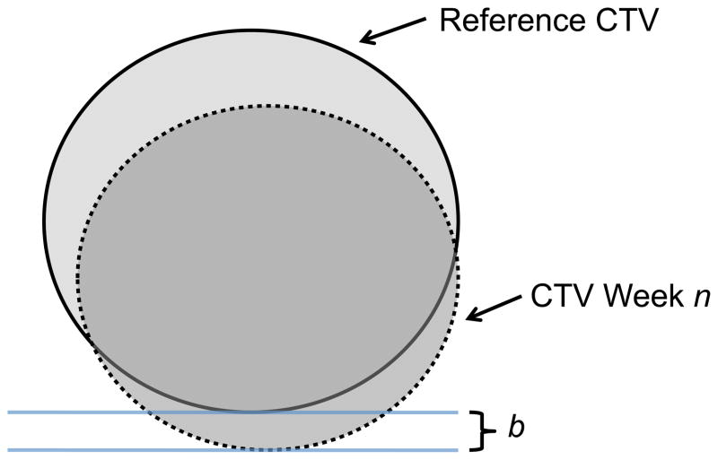
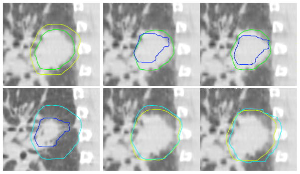
Weekly breath-hold CT scans were acquired in 17 patients undergoing radiotherapy. Deformable registration was used to propagate the GTV and CTV from the first weekly CT image to all other weekly CT images. The on-treatment CT scans were registered rigidly to the planning CT scan based on the GTV location to simulate online guidance, and residual error in the CTV centroids and borders was calculated.
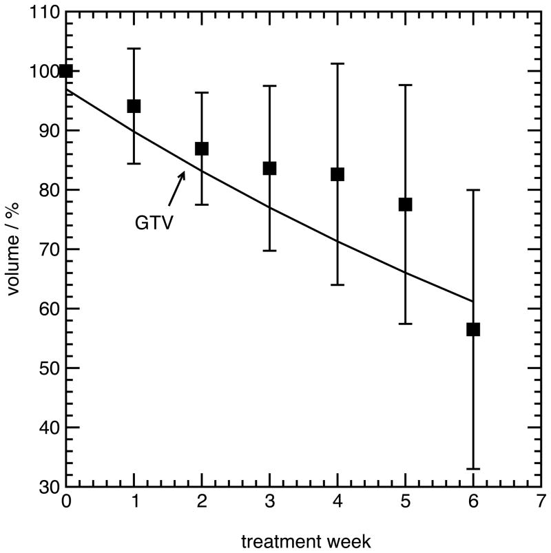
The mean GTV after 5 weeks relative to volume at the beginning of treatment was 77% ± 20%, whereas for the prescribed CTV, it was 92% ± 10%. The mean absolute residual error magnitude in the CTV centroid position after a GTV-based localization was 2.9 ± 3.0 mm, and it varied from 0.3 to 20.0 mm over all patients. Residual error of the CTV centroid was associated with GTV regression and anisotropy of regression during treatment (p = 0.02 and p = 0.03, respectively; Spearman rank correlation). A residual error in CTV border position greater than 2 mm was present in 77% of patients and 50% of fractions. Among these fractions, residual error of the CTV borders was 3.5 ± 1.6 mm (left-right), 3.1 ± 0.9 mm (anterior-posterior), and 6.4 ± 7.5 mm (superior-inferior).
Online guidance based on the visible GTV produces substantial error in CTV localization, particularly for highly regressing tumors. The results of this study will be useful in designing margins for CTV localization or for developing new online CTV localization strategies.
评估在治疗过程中原始定义的临床靶区(CTV)的位置和形状,并评估基于大体肿瘤体积(GTV)的在线计算机断层扫描(CT)引导对 CTV 定位准确性的影响。
对 17 名接受放疗的患者进行每周一次的屏气 CT 扫描。使用可变形配准将 GTV 和 CTV 从第一周的 CT 图像传播到所有其他每周的 CT 图像。基于 GTV 位置将治疗中的 CT 扫描刚性地配准到计划 CT 扫描,以模拟在线引导,并计算 CTV 质心和边界的残余误差。
治疗 5 周后 GTV 相对于治疗开始时的体积为 77%±20%,而对于规定的 CTV,其为 92%±10%。基于 GTV 定位后 CTV 质心位置的平均绝对残余误差幅度为 2.9±3.0mm,所有患者的范围为 0.3-20.0mm。CTV 质心残余误差与 GTV 消退和治疗期间的消退各向异性相关(分别为 p=0.02 和 p=0.03;Spearman 等级相关)。77%的患者和 50%的分次存在 CTV 边界位置的残余误差大于 2mm。在这些分次中,CTV 边界的残余误差为 3.5±1.6mm(左右)、3.1±0.9mm(前后)和 6.4±7.5mm(上下)。
基于可见 GTV 的在线引导会导致 CTV 定位产生大量误差,特别是对于高度消退的肿瘤。本研究的结果将有助于设计 CTV 定位的边界或开发新的在线 CTV 定位策略。