Unit on Neuron-Glia Interactions in Retinal Disease, National Eye Institute, National Institutes of Health, Bethesda, Maryland, United States of America.
PLoS One. 2011 Jan 25;6(1):e15973. doi: 10.1371/journal.pone.0015973.
Microglia represent the primary resident immune cells in the CNS, and have been implicated in the pathology of neurodegenerative diseases. Under basal or "resting" conditions, microglia possess ramified morphologies and exhibit dynamic surveying movements in their processes. Despite the prominence of this phenomenon, the function and regulation of microglial morphology and dynamic behavior are incompletely understood. We investigate here whether and how neurotransmission regulates "resting" microglial morphology and behavior.
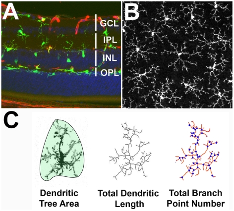
We employed an ex vivo mouse retinal explant system in which endogenous neurotransmission and dynamic microglial behavior are present. We utilized live-cell time-lapse confocal imaging to study the morphology and behavior of GFP-labeled retinal microglia in response to neurotransmitter agonists and antagonists. Patch clamp electrophysiology and immunohistochemical localization of glutamate receptors were also used to investigate direct-versus-indirect effects of neurotransmission by microglia.
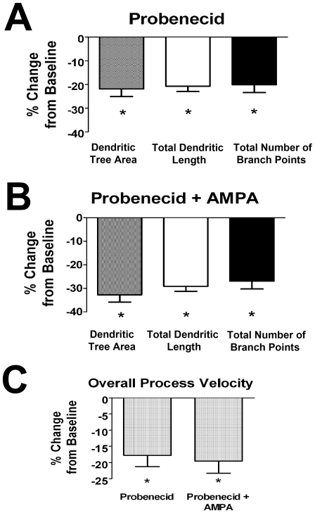
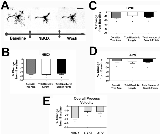
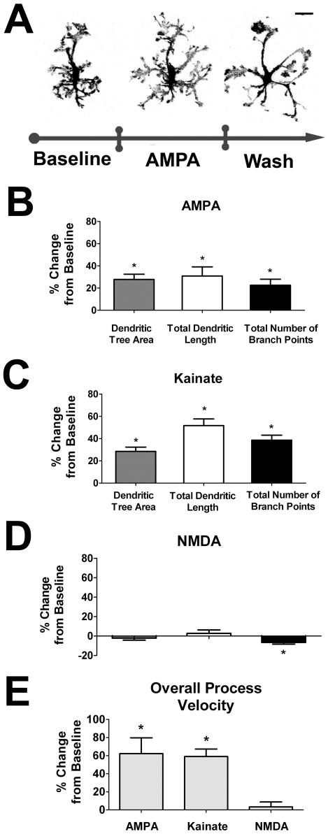
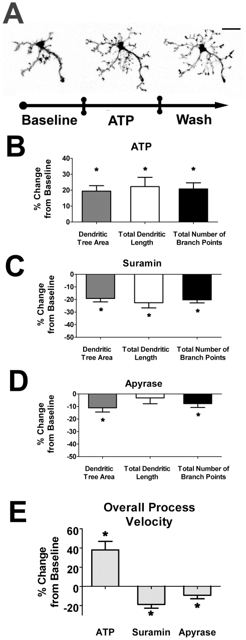
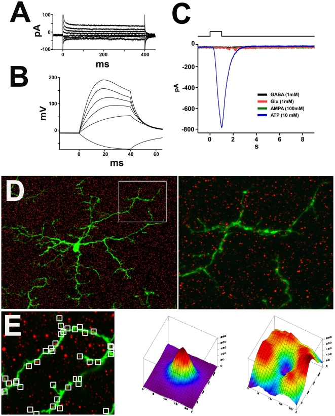
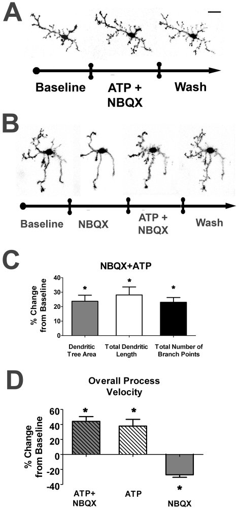
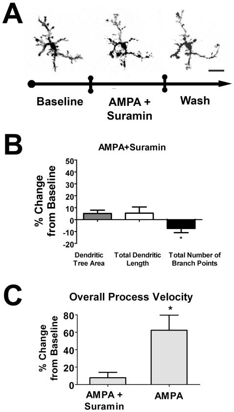
Retinal microglial morphology and dynamic behavior were not cell-autonomously regulated but are instead modulated by endogenous neurotransmission. Morphological parameters and process motility were differentially regulated by different modes of neurotransmission and were increased by ionotropic glutamatergic neurotransmission and decreased by ionotropic GABAergic neurotransmission. These neurotransmitter influences on retinal microglia were however unlikely to be directly mediated; local applications of neurotransmitters were unable to elicit electrical responses on microglia patch-clamp recordings and ionotropic glutamatergic receptors were not located on microglial cell bodies or processes by immunofluorescent labeling. Instead, these influences were mediated indirectly via extracellular ATP, released in response to glutamatergic neurotransmission through probenecid-sensitive pannexin hemichannels.
Our results demonstrate that neurotransmission plays an endogenous role in regulating the morphology and behavior of "resting" microglia in the retina. These findings illustrate a mode of constitutive signaling between the neural and immune compartments of the CNS through which immune cells may be regulated in concert with levels of neural activity.
小胶质细胞是中枢神经系统中主要的常驻免疫细胞,它们与神经退行性疾病的病理学有关。在基础或“静息”状态下,小胶质细胞具有分支状形态,并在其突起中表现出动态探测运动。尽管这一现象很突出,但小胶质细胞形态和动态行为的功能和调节仍不完全清楚。我们在这里研究神经递质是否以及如何调节“静息”小胶质细胞的形态和行为。
我们采用了一种离体小鼠视网膜外植体系统,其中存在内源性神经递质传递和动态小胶质细胞行为。我们利用活细胞延时共焦成像来研究 GFP 标记的视网膜小胶质细胞在神经递质激动剂和拮抗剂作用下的形态和行为。还使用膜片钳电生理学和谷氨酸受体免疫组织化学定位来研究小胶质细胞的神经递质的直接和间接作用。
视网膜小胶质细胞的形态和动态行为不是细胞自主调节的,而是受内源性神经递质调节的。不同的神经递质传递方式对形态参数和突起运动有不同的调节作用,离子型谷氨酸能神经递质传递增加,离子型 GABA 能神经递质传递减少。然而,这些神经递质对视网膜小胶质细胞的影响不太可能是直接介导的;局部应用神经递质不能在小胶质细胞膜片钳记录中引起电反应,离子型谷氨酸能受体也不能通过免疫荧光标记定位在小胶质细胞体或突起上。相反,这些影响是通过细胞外 ATP 的释放间接介导的,ATP 是对谷氨酸能神经递质传递的反应,通过丙磺舒敏感的连接蛋白半通道释放。
我们的结果表明,神经递质在调节视网膜中小胶质细胞的形态和行为方面发挥了内源性作用。这些发现说明了神经和免疫细胞之间通过细胞外 ATP 进行的一种组成性信号传递模式,通过这种模式,免疫细胞可以与神经活动水平协调调节。