Runyan R B, Potts J D, Sharma R V, Loeber C P, Chiang J J, Bhalla R C
Department of Anatomy, University of Iowa, Iowa City 52242.
Cell Regul. 1990 Feb;1(3):301-13. doi: 10.1091/mbc.1.3.301.
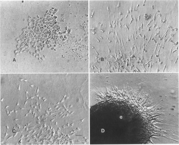
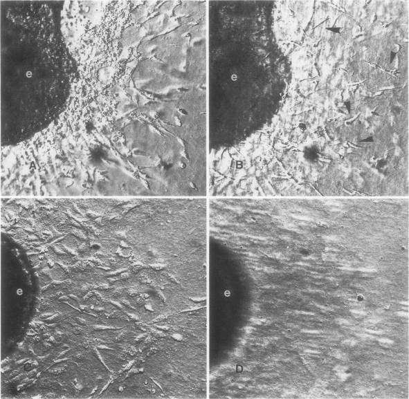
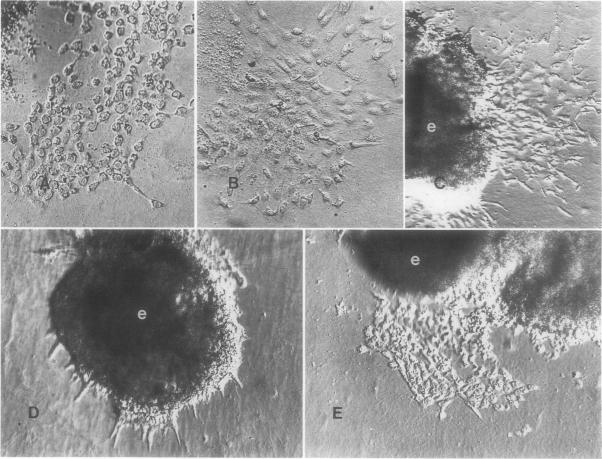
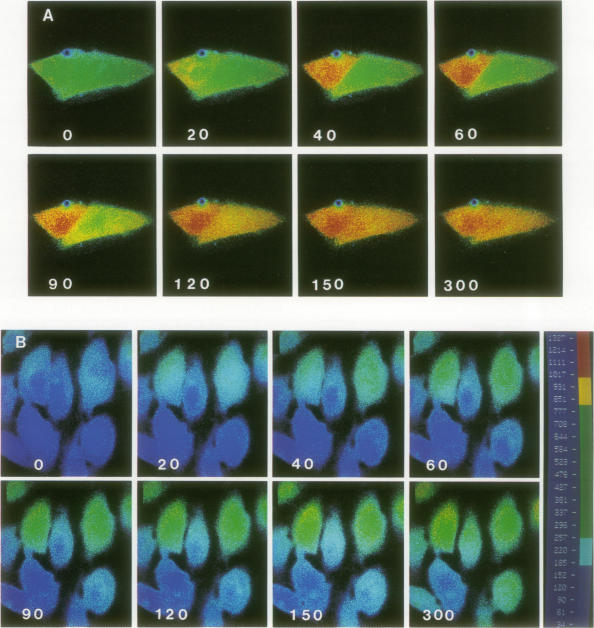
During early cardiac development, progenitors of the valves and septa of the heart are formed by an epithelial-mesenchymal cell transformation of endothelial cells of the atrioventricular (AV) canal. We have previously shown that this event is due to an interaction between the endothelium and products of the myocardium found within the extracellular matrix. The present study examines signal transduction mechanisms governing this differentiation of AV canal endothelium. Activators of protein kinase C (PKC), phorbol myristate acetate (PMA) and mezerein, both produced an incomplete phenotypic transformation of endothelial cells in an in vitro bioassay for transformation. On the other hand, inhibitors of PKC (H-7 and staurosporine) and tyrosine kinase (genistein) blocked cellular transformation in response to the native myocardium or a myocardially-conditioned medium. Intracellular free calcium concentration ([Ca2+]i) was measured in single endothelial cells by microscopic digital analysis of fura 2 fluorescence. Addition of a myocardial conditioned medium containing the transforming stimulus produced a specific increase in [Ca2+]i in "competent" AV canal, but not ventricular, endothelial cells. Epithelial-mesenchymal cell transformation was inhibited by pertussis toxin but not cholera toxin. These data lead to the hypothesis that signal transduction of this tissue interaction is mediated by a G protein and one or more kinase activities. In response to receptor activation, competent AV canal endothelial cells demonstrate an increase in [Ca2+]i. Together, the data provide direct evidence for a regional and temporal regulation of signal transduction processes which mediate a specific extracellular matrix-mediated tissue interaction in the embryo.
在心脏发育早期,心脏瓣膜和间隔的祖细胞由房室(AV)管内皮细胞的上皮-间充质细胞转化形成。我们之前已经表明,这一事件是由于内皮细胞与细胞外基质中发现的心肌产物之间的相互作用所致。本研究考察了调控AV管内皮细胞这种分化的信号转导机制。蛋白激酶C(PKC)的激活剂佛波酯肉豆蔻酸酯(PMA)和大戟二萜醇在体外转化生物测定中均使内皮细胞产生不完全的表型转化。另一方面,PKC抑制剂(H-7和星形孢菌素)和酪氨酸激酶抑制剂(染料木黄酮)可阻断对天然心肌或心肌条件培养基的细胞转化反应。通过fura 2荧光的显微镜数字分析测量单个内皮细胞内的游离钙浓度([Ca2+]i)。添加含有转化刺激物的心肌条件培养基会使“有反应能力的”AV管而非心室的内皮细胞中的[Ca2+]i特异性升高。上皮-间充质细胞转化受到百日咳毒素的抑制,但不受霍乱毒素的抑制。这些数据得出这样的假说,即这种组织相互作用的信号转导由一种G蛋白和一种或多种激酶活性介导。响应受体激活,有反应能力的AV管内皮细胞表现出[Ca2+]i升高。总之,这些数据为信号转导过程的区域和时间调控提供了直接证据,该调控介导了胚胎中一种特定的细胞外基质介导的组织相互作用。