• 文献检索
  • 文档翻译
  • 深度研究
  • 学术资讯
  • Suppr Zotero 插件Zotero 插件
  • 邀请有礼
  • 套餐&价格
  • 历史记录
应用&插件
Suppr Zotero 插件Zotero 插件浏览器插件Mac 客户端Windows 客户端微信小程序
定价
高级版会员购买积分包购买API积分包
服务
文献检索文档翻译深度研究API 文档MCP 服务
关于我们
关于 Suppr公司介绍联系我们用户协议隐私条款
关注我们

Suppr 超能文献

核心技术专利:CN118964589B侵权必究
粤ICP备2023148730 号-1Suppr @ 2026

文献检索

告别复杂PubMed语法,用中文像聊天一样搜索,搜遍4000万医学文献。AI智能推荐,让科研检索更轻松。

立即免费搜索

文件翻译

保留排版,准确专业,支持PDF/Word/PPT等文件格式,支持 12+语言互译。

免费翻译文档

深度研究

AI帮你快速写综述,25分钟生成高质量综述,智能提取关键信息,辅助科研写作。

立即免费体验

钙/NFAT 信号促进早期肾发生。

Calcium/NFAT signalling promotes early nephrogenesis.

机构信息

MRC Human Genetics Unit, Institute of Genetics & Molecular Medicine, Western General Hospital, Crewe Road, Edinburgh EH4 2XU, UK.

出版信息

Dev Biol. 2011 Apr 15;352(2):288-98. doi: 10.1016/j.ydbio.2011.01.033. Epub 2011 Feb 3.

DOI:10.1016/j.ydbio.2011.01.033
PMID:21295565
原文链接:https://pmc.ncbi.nlm.nih.gov/articles/PMC3070816/
Abstract

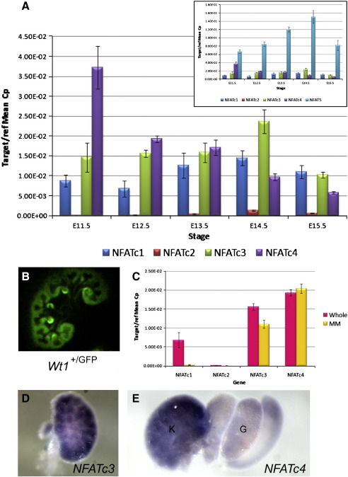
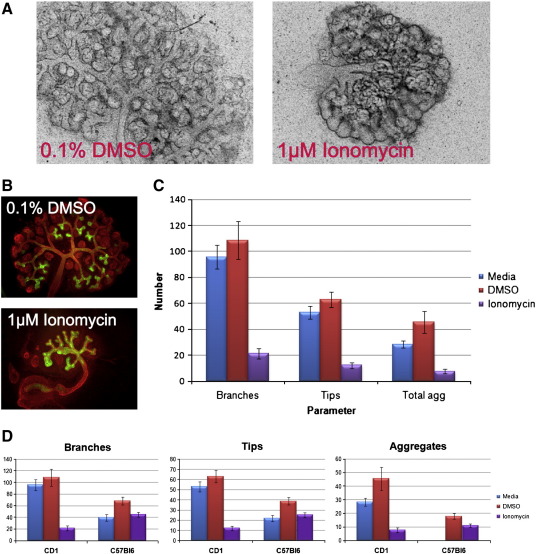
A number of Wnt genes are expressed during, and are known to be essential for, early kidney development. It is typically assumed that their products will act through the canonical β-catenin signalling pathway. We have found evidence that suggests canonical Wnt signalling is not active in the early nephrogenic metanephric mesenchyme, but instead provide expressional and functional evidence that implicates the non-canonical Calcium/NFAT Wnt signalling pathway in nephrogenesis. Members of the NFAT (Nuclear Factor Activated in T cells) transcription factor gene family are expressed throughout murine kidney morphogenesis and NFATc3 is localised to the developing nephrons. Treatment of kidney rudiments with Cyclosporin A (CSA), an inhibitor of Calcium/NFAT signalling, decreases nephron formation--a phenotype similar to that in Wnt4(-/-) embryos. Treatment of Wnt4(-/-) kidneys with Ionomycin, an activator of the pathway, partially rescues the phenotype. We propose that the non-canonical Calcium/NFAT Wnt signalling pathway plays an important role in early mammalian renal development and is required for complete MET during nephrogenesis, potentially acting downstream of Wnt4.

摘要

许多 Wnt 基因在肾脏早期发育过程中表达,并被认为对其至关重要。通常假设它们的产物将通过经典的β-连环蛋白信号通路发挥作用。我们已经发现证据表明,经典 Wnt 信号通路在早期肾原基的中肾间充质中不活跃,但相反,提供表达和功能证据表明非经典钙/ NFAT Wnt 信号通路在肾发生中起作用。NFAT(T 细胞中激活的核因子)转录因子基因家族的成员在整个鼠肾形态发生过程中表达,NFATc3 定位于发育中的肾单位。用环孢素 A(CSA)处理肾原基,这是一种钙/NFAT 信号通路的抑制剂,会减少肾单位的形成 - 这种表型类似于 Wnt4(-/-)胚胎的表型。用离子霉素处理 Wnt4(-/-)肾脏,这是该途径的激活剂,部分挽救了表型。我们提出,非经典钙/ NFAT Wnt 信号通路在哺乳动物肾脏早期发育中起重要作用,并且在肾发生过程中需要完全的 MET,可能在 Wnt4 下游发挥作用。

https://cdn.ncbi.nlm.nih.gov/pmc/blobs/28a4/3070816/dd7291632038/gr7.jpg
https://cdn.ncbi.nlm.nih.gov/pmc/blobs/28a4/3070816/b0168e03c690/gr8.jpg
https://cdn.ncbi.nlm.nih.gov/pmc/blobs/28a4/3070816/456c929a5f11/gr9.jpg
https://cdn.ncbi.nlm.nih.gov/pmc/blobs/28a4/3070816/9f6bfc050933/gr1.jpg
https://cdn.ncbi.nlm.nih.gov/pmc/blobs/28a4/3070816/a4ac7591a7f1/gr2.jpg
https://cdn.ncbi.nlm.nih.gov/pmc/blobs/28a4/3070816/8d60ae8ea31f/gr3.jpg
https://cdn.ncbi.nlm.nih.gov/pmc/blobs/28a4/3070816/149e458f93ea/gr4.jpg
https://cdn.ncbi.nlm.nih.gov/pmc/blobs/28a4/3070816/0f3475f1e199/gr5.jpg
https://cdn.ncbi.nlm.nih.gov/pmc/blobs/28a4/3070816/411f38c352d7/gr6.jpg
https://cdn.ncbi.nlm.nih.gov/pmc/blobs/28a4/3070816/dd7291632038/gr7.jpg

相似文献

1
Calcium/NFAT signalling promotes early nephrogenesis.钙/NFAT 信号促进早期肾发生。
Dev Biol. 2011 Apr 15;352(2):288-98. doi: 10.1016/j.ydbio.2011.01.033. Epub 2011 Feb 3.
2
Wnt4 induces nephronic tubules in metanephric mesenchyme by a non-canonical mechanism.Wnt4 通过非经典机制诱导后肾间充质形成肾单位。
Dev Biol. 2011 Apr 1;352(1):58-69. doi: 10.1016/j.ydbio.2011.01.012. Epub 2011 Jan 21.
3
Canonical WNT signaling during kidney development.肾脏发育过程中的经典WNT信号传导。
Am J Physiol Renal Physiol. 2007 Aug;293(2):F494-500. doi: 10.1152/ajprenal.00416.2006. Epub 2007 May 9.
4
Control of kidney development by calcium ions.钙离子对肾脏发育的控制。
Biochimie. 2011 Dec;93(12):2126-31. doi: 10.1016/j.biochi.2011.07.007. Epub 2011 Jul 23.
5
Wnt/beta-catenin signaling regulates nephron induction during mouse kidney development.Wnt/β-连环蛋白信号通路在小鼠肾脏发育过程中调节肾单位诱导。
Development. 2007 Jul;134(13):2533-9. doi: 10.1242/dev.006155. Epub 2007 May 30.
6
Nuclear factor of activated T cells (NFAT) proteins repress canonical Wnt signaling via its interaction with Dishevelled (Dvl) protein and participate in regulating neural progenitor cell proliferation and differentiation.激活 T 细胞的核因子 (NFAT) 蛋白通过与 Dvl 蛋白相互作用抑制经典 Wnt 信号通路,并参与调节神经前体细胞的增殖和分化。
J Biol Chem. 2011 Oct 28;286(43):37399-405. doi: 10.1074/jbc.M111.251165. Epub 2011 Aug 31.
7
Cyclosporin A promotes growth and invasiveness in vitro of human first-trimester trophoblast cells via MAPK3/MAPK1-mediated AP1 and Ca2+/calcineurin/NFAT signaling pathways.环孢素A通过MAPK3/MAPK1介导的AP1和Ca2+/钙调神经磷酸酶/NFAT信号通路促进人早孕滋养层细胞的体外生长和侵袭。
Biol Reprod. 2008 Jun;78(6):1102-10. doi: 10.1095/biolreprod.107.063503. Epub 2008 Mar 5.
8
R-spondin signalling is essential for the maintenance and differentiation of mouse nephron progenitors.R-spondin 信号对于维持和分化小鼠肾祖细胞至关重要。
Elife. 2020 May 1;9:e53895. doi: 10.7554/eLife.53895.
9
Essential role of nuclear factor of activated T cells (NFAT)-mediated Wnt signaling in osteoblast differentiation induced by strontium ranelate.核因子活化 T 细胞(NFAT)介导的 Wnt 信号通路在雷奈酸锶诱导的成骨细胞分化中的重要作用。
J Biol Chem. 2010 Aug 13;285(33):25251-8. doi: 10.1074/jbc.M110.110502. Epub 2010 Jun 16.
10
WNT/beta-catenin signaling in nephron progenitors and their epithelial progeny.肾单位祖细胞及其上皮后代中的WNT/β-连环蛋白信号传导
Kidney Int. 2008 Oct;74(8):1004-8. doi: 10.1038/ki.2008.322. Epub 2008 Jul 16.

引用本文的文献

1
Deciphering the cellular and molecular landscapes of Wnt/β-catenin signaling in mouse embryonic kidney development.解析小鼠胚胎肾发育过程中Wnt/β-连环蛋白信号通路的细胞和分子图景。
Comput Struct Biotechnol J. 2024 Sep 2;23:3368-3378. doi: 10.1016/j.csbj.2024.08.025. eCollection 2024 Dec.
2
3D Hydrogel Encapsulation Regulates Nephrogenesis in Kidney Organoids.3D 水凝胶包封调控肾类器官中的肾发生。
Adv Mater. 2024 Apr;36(14):e2308325. doi: 10.1002/adma.202308325. Epub 2024 Jan 10.
3
Principles of human and mouse nephron development.

本文引用的文献

1
Wnt4 induces nephronic tubules in metanephric mesenchyme by a non-canonical mechanism.Wnt4 通过非经典机制诱导后肾间充质形成肾单位。
Dev Biol. 2011 Apr 1;352(1):58-69. doi: 10.1016/j.ydbio.2011.01.012. Epub 2011 Jan 21.
2
A sensitive and bright single-cell resolution live imaging reporter of Wnt/ß-catenin signaling in the mouse.一种灵敏且清晰的小鼠Wnt/β-连环蛋白信号通路的单细胞分辨率活体成像报告基因。
BMC Dev Biol. 2010 Dec 21;10:121. doi: 10.1186/1471-213X-10-121.
3
Activation of NFAT signaling in podocytes causes glomerulosclerosis.
人类和小鼠肾单位发育的原理。
Nat Rev Nephrol. 2022 Oct;18(10):628-642. doi: 10.1038/s41581-022-00598-5. Epub 2022 Jul 22.
4
The complex role of Wnt ligands in type 2 diabetes mellitus and related complications.Wnt 配体在 2 型糖尿病及其相关并发症中的复杂作用。
J Cell Mol Med. 2021 Jul;25(14):6479-6495. doi: 10.1111/jcmm.16663. Epub 2021 May 27.
5
WNT5B in Physiology and Disease.WNT5B在生理与疾病中的作用
Front Cell Dev Biol. 2021 May 4;9:667581. doi: 10.3389/fcell.2021.667581. eCollection 2021.
6
Embryonic Kidney Development, Stem Cells and the Origin of Wilms Tumor.胚胎肾发育、干细胞与肾母细胞瘤的起源。
Genes (Basel). 2021 Feb 23;12(2):318. doi: 10.3390/genes12020318.
7
Inhibition of GSK3 Represses the Expression of Retinoic Acid Synthetic Enzyme ALDH1A2 via Wnt/β-Catenin Signaling in WiT49 Cells.在WiT49细胞中,GSK3的抑制通过Wnt/β-连环蛋白信号通路抑制视黄酸合成酶ALDH1A2的表达。
Front Cell Dev Biol. 2020 Mar 17;8:94. doi: 10.3389/fcell.2020.00094. eCollection 2020.
8
Non-canonical Wnt signalling regulates scarring in biliary disease via the planar cell polarity receptors.非经典 Wnt 信号通路通过平面细胞极性受体调节胆道疾病的瘢痕形成。
Nat Commun. 2020 Jan 23;11(1):445. doi: 10.1038/s41467-020-14283-3.
9
Stem cell-derived kidney organoids: engineering the vasculature.干细胞衍生的肾脏类器官:血管工程。
Cell Mol Life Sci. 2020 Jun;77(12):2257-2273. doi: 10.1007/s00018-019-03401-0. Epub 2019 Dec 5.
10
NHA2 promotes cyst development in an in vitro model of polycystic kidney disease.NHA2 促进了多囊肾病体外模型中的囊肿发育。
J Physiol. 2019 Jan;597(2):499-519. doi: 10.1113/JP276796. Epub 2018 Oct 17.
NFAT 信号通路在足细胞中的激活导致肾小球硬化。
J Am Soc Nephrol. 2010 Oct;21(10):1657-66. doi: 10.1681/ASN.2009121253. Epub 2010 Jul 22.
4
Regulation of embryonic kidney branching morphogenesis and glomerular development by KISS1 receptor (Gpr54) through NFAT2- and Sp1-mediated Bmp7 expression.通过 NFAT2 和 Sp1 介导的 Bmp7 表达, Kiss1 受体(Gpr54)调控胚胎肾分支形态发生和肾小球发育。
J Biol Chem. 2010 Jun 4;285(23):17811-20. doi: 10.1074/jbc.M110.130740. Epub 2010 Apr 7.
5
High-resolution gene expression analysis of the developing mouse kidney defines novel cellular compartments within the nephron progenitor population.对发育中的小鼠肾脏进行高分辨率基因表达分析,确定了肾祖细胞群体中的新型细胞区室。
Dev Biol. 2009 Sep 15;333(2):312-23. doi: 10.1016/j.ydbio.2009.06.043. Epub 2009 Jul 8.
6
Wnt9b signaling regulates planar cell polarity and kidney tubule morphogenesis.Wnt9b信号传导调节平面细胞极性和肾小管形态发生。
Nat Genet. 2009 Jul;41(7):793-9. doi: 10.1038/ng.400. Epub 2009 Jun 21.
7
Requirement of Wnt/beta-catenin signaling in pronephric kidney development.原肾发育过程中Wnt/β-连环蛋白信号通路的需求。
Mech Dev. 2009 Mar-Apr;126(3-4):142-59. doi: 10.1016/j.mod.2008.11.007. Epub 2008 Dec 7.
8
A genecentric Human Protein Atlas for expression profiles based on antibodies.基于抗体的、以基因为中心的人类蛋白质图谱用于表达谱分析。
Mol Cell Proteomics. 2008 Oct;7(10):2019-27. doi: 10.1074/mcp.R800013-MCP200.
9
An increase in intracellular Ca2+ is involved in pronephric tubule differentiation in the amphibian Xenopus laevis.细胞内钙离子浓度的升高参与了两栖动物非洲爪蟾原肾管的分化过程。
Dev Biol. 2008 Sep 15;321(2):357-67. doi: 10.1016/j.ydbio.2008.06.029. Epub 2008 Jul 2.
10
Canonical WNT/beta-catenin signaling is required for ureteric branching.经典WNT/β-连环蛋白信号通路是输尿管分支所必需的。
Dev Biol. 2008 May 1;317(1):83-94. doi: 10.1016/j.ydbio.2008.02.010. Epub 2008 Feb 21.