Sen Shamik, Kumar Sanjay
Department of Bioengineering, University of California, Berkeley, Berkeley, CA 94720-1762 USA.
Cell Mol Bioeng. 2009 Jun;2(2):218-230. doi: 10.1007/s12195-009-0057-7. Epub 2009 May 5.
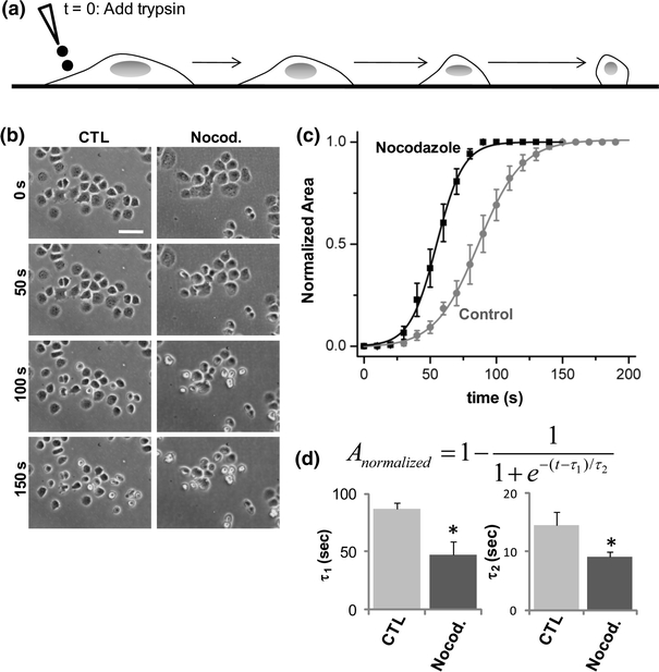
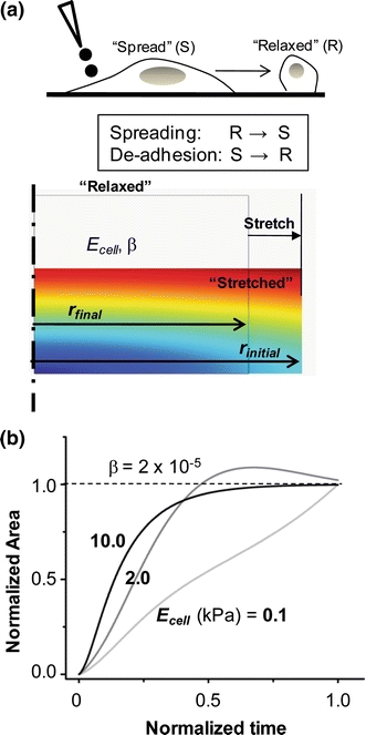
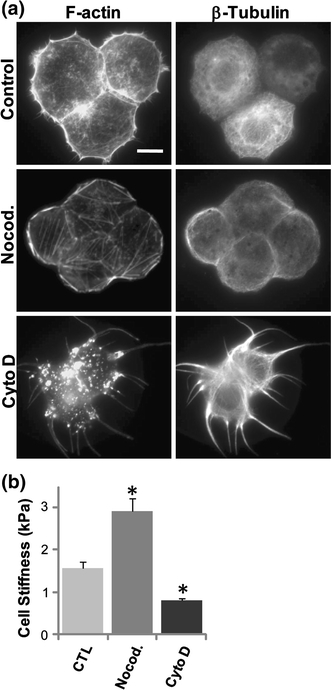
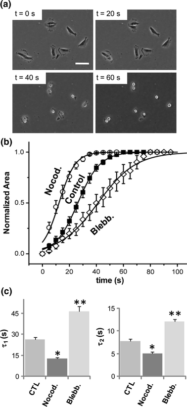
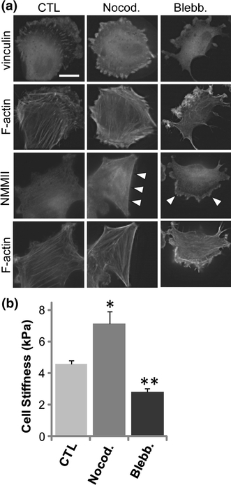
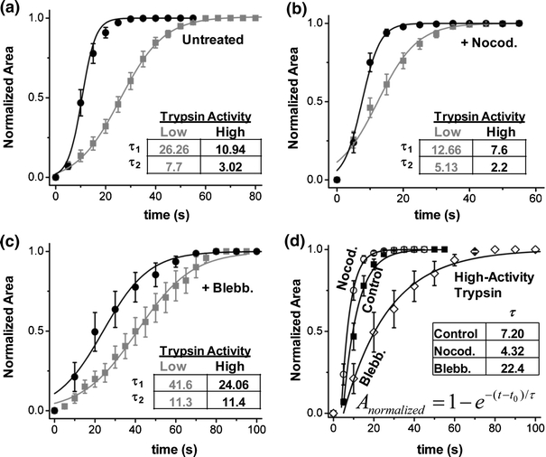
Measurement of the mechanical properties of single cells is of increasing interest both from a fundamental cell biological perspective and in the context of disease diagnostics. In this study, we show that tracking cell shape dynamics during trypsin-induced de-adhesion can serve as a simple but extremely useful tool for probing the contractility of adherent cells. When treated with trypsin, both SW13(-/-) epithelial cells and U373 MG glioma cells exhibit a brief lag period followed by a concerted retraction to a rounded shape. The time-response of the normalized cell area can be fit to a sigmoidal curve with two characteristic time constants that rise and fall when cells are treated with blebbistatin and nocodazole, respectively. These differences can be attributed to actomyosin-based cytoskeletal remodeling, as evidenced by the prominent buildup of stress fibers in nocodazole-treated SW13(-/-) cells, which are also two-fold stiffer than untreated cells. Similar results observed in U373 MG cells highlights the direct association between cell stiffness and the de-adhesion response. Faster de-adhesion is obtained with higher trypsin concentration, with nocodazole treatment further expediting the process and blebbistatin treatment blunting the response. A simple finite element model confirms that faster contraction is achieved with increased stiffness.
从基础细胞生物学角度以及疾病诊断的背景来看,单细胞力学特性的测量越来越受到关注。在本研究中,我们表明,在胰蛋白酶诱导的去黏附过程中跟踪细胞形状动态变化,可作为一种简单但极其有用的工具,用于探究贴壁细胞的收缩性。用胰蛋白酶处理时,SW13(-/-)上皮细胞和U373 MG胶质瘤细胞均表现出短暂的延迟期,随后协同收缩成圆形。归一化细胞面积的时间响应可以拟合为一条S形曲线,有两个特征时间常数,分别在用blebbistatin和诺考达唑处理细胞时上升和下降。这些差异可归因于基于肌动球蛋白的细胞骨架重塑,在用诺考达唑处理的SW13(-/-)细胞中应力纤维的显著积累证明了这一点,这些细胞的硬度也比未处理细胞高两倍。在U373 MG细胞中观察到的类似结果突出了细胞硬度与去黏附反应之间的直接关联。胰蛋白酶浓度越高,去黏附速度越快,诺考达唑处理进一步加快了这一过程,而blebbistatin处理则减弱了反应。一个简单的有限元模型证实,随着硬度增加,收缩速度加快。