Ghoubay-Benallaoua D, Basli E, Goldschmidt P, Pecha F, Chaumeil C, Laroche L, Borderie V
Institut de la Vision, UPMC Univ Paris 06, UMR_S 968 / INSERM, U968 / CHNO des XV-XX / CNRS, UMR_7210, Paris, France.
Mol Vis. 2011 Feb 1;17:341-54.
To study the kinetics of growth and the phenotype of cells cultured from human limbal explants in a cholera toxin-free medium with no feeder cell layer.
Human organ-cultured corneas were used to prepare limbal explants (full-thickness and superficial limbal explants) and corneal stromal explants. Cell growth kinetics and phenotypes were assessed by cultivating explants in cholera toxin-free Green medium. Epithelial and progenitor cell markers were assessed by immunocytochemistry, flow cytometry, and Reverse Transcription and Polymerase Chain Reaction (RT-PCR).
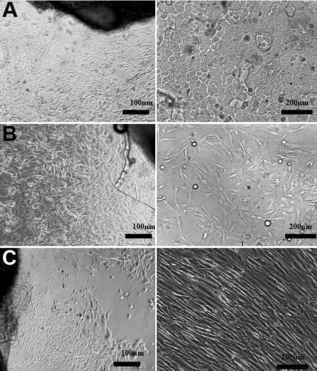
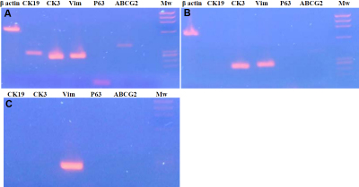
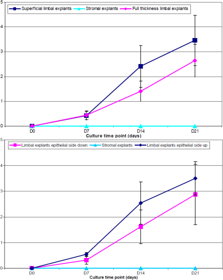
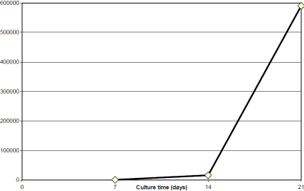
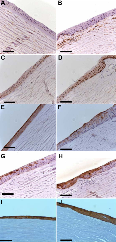
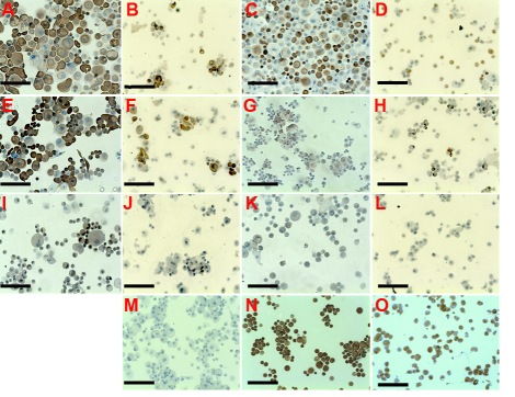
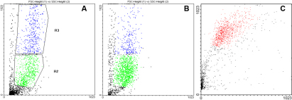
The successful epithelial cell growth rates from full thickness limbal explant and superficial limbal explant tissues were 41 and 86%, respectively (p=0.0001). The mean cell area and the percentage of small cells in superficial and full-thickness explant cultures were, respectively, 317 µm(2) and 429 µm(2), and 8.9% and 1.7% (p<0.001). The percentage of positive cells in superficial and full-thickness limbal explant cultures as assessed by immunocytochemistry were the following: broad spectrum cytokeratins (cytokeratins 4, 5, 6, 8, 10, 13, and 18 [MNF116]), 82%/37% (p=0.01); cytokeratin 3 (CK3), 74%/25% (p=0.009); cytokeratin 19 (CK19), 46%/25% (p=0.19); vimentin, 56%/53% (p=0.48); delta N p63α, 54%/0% (p<0.001); and ABCG2, 5%/0% (p=0.1). Flow cytometry showed a higher percentage of small cells, a higher percentage of MNF116+ cells, and stronger expression of progenitor-associated markers in superficial than in full-thickness explant cultures. For superficial limbal explant cultures, analysis of the expression profiles for various mRNAs at the end of 21 days of culture showed high levels of expression of the mRNAs encoding CK3, vimentin, and CK19. The expression of mRNA of delta N p63α and ABCG2 was weaker. Cultures obtained from full-thickness limbal explants featured no expression of mRNA of CK19, delta N p63α, and ABCG2, whereas mRNAs encoding CK3 and vimentin were detected. Human corneal stromal explants cultured with the same medium featured late cell growth, large mean cell area (2,529 µm(2)), no expression of cytokeratins, delta N p63α, and ABCG2, and high expression of vimentin.
Superficial limbal explants appear to be superior to full-thickness limbal explants for growing human limbal epithelial cells. Preparation of explants using surgical facilities (i.e., operating microscope and microsurgical blades) led to a dramatic increase in the percentage of successful cultures, higher epithelial cell growth, decreased fibroblast contamination, and better preservation of limbal epithelial progenitors.
研究在无霍乱毒素且无饲养细胞层的培养基中,从人角膜缘外植体培养的细胞的生长动力学和表型。
用人器官培养的角膜制备角膜缘外植体(全层和浅层角膜缘外植体)和角膜基质外植体。通过在无霍乱毒素的绿色培养基中培养外植体来评估细胞生长动力学和表型。通过免疫细胞化学、流式细胞术以及逆转录聚合酶链反应(RT-PCR)评估上皮细胞和祖细胞标志物。
全层角膜缘外植体和浅层角膜缘外植体组织上皮细胞的成功生长率分别为41%和86%(p = 0.0001)。浅层和全层外植体培养物中小细胞的平均细胞面积和百分比分别为317 µm²和429 µm²,以及8.9%和1.7%(p < 0.001)。通过免疫细胞化学评估,浅层和全层角膜缘外植体培养物中阳性细胞的百分比情况如下:广谱细胞角蛋白(细胞角蛋白4、5、6、8、10、13和18 [MNF116]),82%/37%(p = 0.01);细胞角蛋白3(CK3),74%/25%(p = 0.009);细胞角蛋白19(CK19),46%/25%(p = 0.19);波形蛋白,56%/53%(p = 0.48);ΔN p63α,54%/0%(p < 0.001);以及ABCG2,5%/0%(p = 0.1)。流式细胞术显示,与全层外植体培养物相比,浅层培养物中小细胞的百分比更高、MNF116 +细胞的百分比更高,并且祖细胞相关标志物的表达更强。对于浅层角膜缘外植体培养物,在培养21天结束时对各种mRNA表达谱的分析显示,编码CK3、波形蛋白和CK19的mRNA表达水平较高。ΔN p63α和ABCG2的mRNA表达较弱。从全层角膜缘外植体获得的培养物中未检测到CK19、ΔN p63α和ABCG2的mRNA表达,而检测到编码CK3和波形蛋白的mRNA。用相同培养基培养的人角膜基质外植体细胞生长较晚,平均细胞面积大(2529 µm²),未表达细胞角蛋白、ΔN p63α和ABCG2,波形蛋白表达高。
对于培养人角膜缘上皮细胞,浅层角膜缘外植体似乎优于全层角膜缘外植体。使用手术设备(即手术显微镜和显微外科刀片)制备外植体导致成功培养的百分比显著增加、上皮细胞生长更高、成纤维细胞污染减少以及角膜缘上皮祖细胞的保存更好。