Department of Neuroscience, University of Connecticut Health Center, Farmington, CT, USA.
Stroke. 2011 Apr;42(4):1090-6. doi: 10.1161/STROKEAHA.110.594861. Epub 2011 Feb 10.
Emerging data suggest that the molecular cell death pathways triggered by ischemic insults differ in the male and female brain. Cell death in males is initiated by poly(ADP-ribose) polymerase-1 (PARP-1) activation; however, manipulation of this pathway paradoxically increases ischemic damage in females. In contrast, females are exquisitely sensitive to caspase-mediated cell death. The effect of caspase inhibition in PARP-1 knockout mice was evaluated to determine if the detrimental effects of PARP deletion in females were secondary to increased caspase activation.
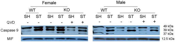
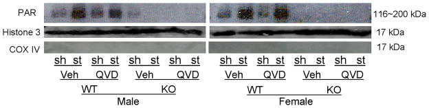
Focal stroke was induced by transient or permanent middle cerebral artery occlusion (MCAO) in wild-type (WT) and PARP-1(-/-) mice of both sexes. The pan-caspase inhibitor, quinoline-Val-Asp(Ome)-CH2-O-phenoxy (Q-VD-OPh), was administered 90 minutes after middle cerebral artery occlusion. Infarct size and neurological sores were assessed. Separate cohorts were used for protein analysis for PAR, Apoptosis inducing factor (AIF), caspase-9, and caspase-3.
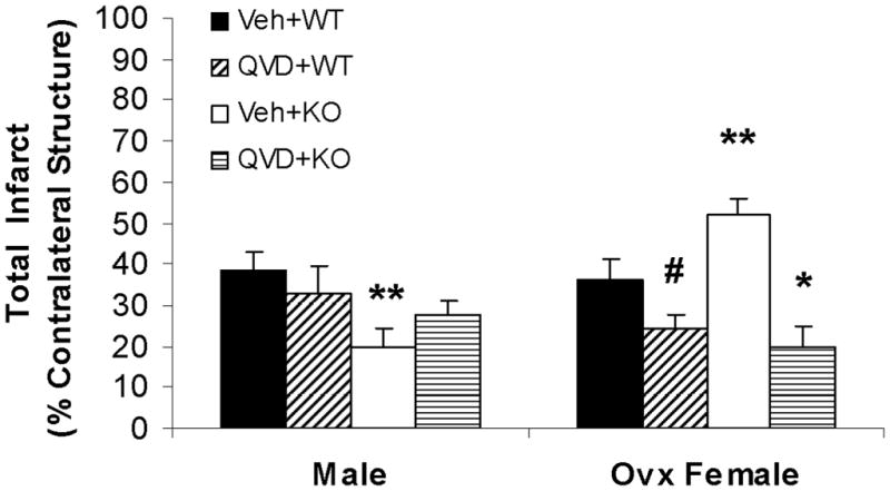
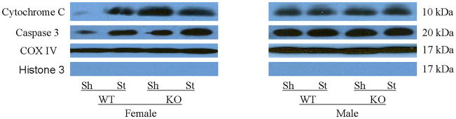
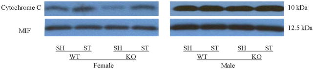
WT mice of both sexes had increased nuclear AIF after stroke compared to PARP-1(-/-) mice. PARP-1(-/-) females had higher mitochondrial cytochrome C and activated caspase-9 and -3 levels than WT female mice. PARP-1(-/-) females also had an increase in stroke-induced cytosolic cytochrome C release compared with WT females, which was not seen in males. Q-VD-OPh decreased caspase-9 in both males and females but only led to reduction of infarct in females. PARP-1(-/-) males had smaller infarcts, whereas PARP-1(-/-) females had larger strokes compared with WT. Q-VD-OPh significantly decreased infarct in both WT and PARP-1(-/-) females in both transient and permanent MCAO models, but had no effect in males.
Deletion of PARP-1 reduces infarct in males but exacerbates injury in females. PARP-1(-/-) females have enhanced caspase activation. The detrimental effects of PARP loss in females can be reversed with caspase inhibition.
新出现的数据表明,在男性和女性大脑中,由缺血性损伤引发的分子细胞死亡途径不同。在男性中,细胞死亡是由多聚(ADP-核糖)聚合酶-1(PARP-1)的激活引发的;然而,对该途径的操作却反常地增加了女性的缺血性损伤。相比之下,女性对半胱氨酸天冬氨酸蛋白酶介导的细胞死亡非常敏感。评估了半胱氨酸天冬氨酸蛋白酶抑制剂对 PARP-1 基因敲除小鼠的影响,以确定 PARP 缺失对女性的有害影响是否是由于半胱氨酸天冬氨酸蛋白酶的激活增加所致。
通过短暂或永久性大脑中动脉闭塞(MCAO)在雄性和雌性野生型(WT)和 PARP-1(-/-)小鼠中诱导局灶性中风。在大脑中动脉闭塞后 90 分钟给予泛半胱氨酸天冬氨酸蛋白酶抑制剂 quinoline-Val-Asp(Ome)-CH2-O-phenoxy(Q-VD-OPh)。评估梗死面积和神经评分。使用单独的队列进行 PAR、凋亡诱导因子(AIF)、半胱氨酸天冬氨酸蛋白酶-9 和 -3 的蛋白分析。
与 PARP-1(-/-)小鼠相比,雄性和雌性 WT 小鼠中风后核 AIF 增加。PARP-1(-/-)雌性小鼠的线粒体细胞色素 C 和激活的半胱氨酸天冬氨酸蛋白酶-9 和 -3 水平高于 WT 雌性小鼠。PARP-1(-/-)雌性小鼠中风后细胞质细胞色素 C 释放增加,而雄性小鼠则没有。Q-VD-OPh 降低了雄性和雌性小鼠的半胱氨酸天冬氨酸蛋白酶-9,但仅导致雌性小鼠的梗死面积减小。与 WT 相比,PARP-1(-/-)雄性小鼠的梗死面积较小,而 PARP-1(-/-)雌性小鼠的中风较大。Q-VD-OPh 显著降低了两种 WT 和 PARP-1(-/-)雌性小鼠在短暂和永久性 MCAO 模型中的梗死面积,但对雄性小鼠没有影响。
PARP-1 的缺失减少了雄性小鼠的梗死,但加剧了雌性小鼠的损伤。PARP-1(-/-)雌性小鼠半胱氨酸天冬氨酸蛋白酶的激活增强。用半胱氨酸天冬氨酸蛋白酶抑制剂可以逆转 PARP 缺失对女性的有害影响。