Veda P, Srinivasaiah M
Department of Pathology, India.
J Lab Physicians. 2010 Jul;2(2):117-20. doi: 10.4103/0974-2727.72216.
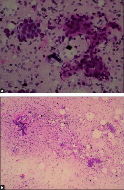
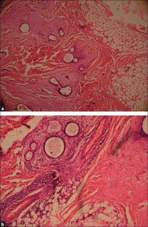
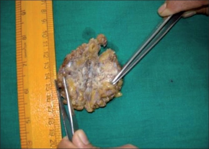
Incisional endometriosis (IE) is a rare entity reported in 0.03-1.08% of women following obstetric or gynecologic surgeries. Most cases reported in literature have appeared after cesarean sections and were often clinically mistaken for hernia, abscess, suture granuloma or lipoma. We hereby report a case of IE following a second trimester hysterotomy, which was diagnosed by fine needle aspiration cytology (FNAC). Our patient was 26 years old, presenting with a mass over anterior abdominal wall, associated with incapacitating pain during each menstrual cycle. FNAC showed epithelial cells, stromal cells and hemosiderin laden macrophages. Based on the typical history, clinical and cytological features, the diagnosis of IE was established. Wide surgical excision was done and the resulting rectus sheath defect was repaired. Patient was followed for 6 months during which time she was symptom free. This article also reviews the spectrum of cytological features and the rare possibility of malignant transformation that can occur in IE.
切口子宫内膜异位症(IE)是一种罕见疾病,在0.03%-1.08%的妇产科手术后女性中被报道。文献中报道的大多数病例出现在剖宫产术后,临床上常被误诊为疝气、脓肿、缝线肉芽肿或脂肪瘤。我们在此报告一例孕中期子宫切开术后发生的IE病例,该病例通过细针穿刺抽吸细胞学检查(FNAC)得以确诊。我们的患者为26岁女性,前腹壁出现肿物,每个月经周期伴有剧痛。FNAC显示有上皮细胞、间质细胞和含铁血黄素巨噬细胞。基于典型病史、临床及细胞学特征,确诊为IE。进行了广泛手术切除,并修复了由此产生的腹直肌鞘缺损。对患者随访6个月,在此期间她无症状。本文还回顾了IE的细胞学特征谱以及可能发生的罕见恶性转化情况。