Gene Therapy Program, Department of Cellular Biology, LSU Health Sciences Center, Shreveport, Louisiana, United States of America.
PLoS One. 2011 Feb 9;6(2):e16792. doi: 10.1371/journal.pone.0016792.
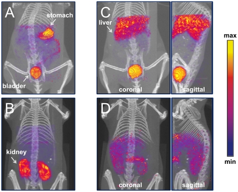
As the limits of existing treatments for cancer are recognized, clearly novel therapies must be considered for successful treatment; cancer therapy using adenovirus vectors is a promising strategy. However tracking the biodistribution of adenovirus vectors in vivo is limited to invasive procedures such as biopsies, which are error prone, non-quantitative, and do not give a full representation of the pharmacokinetics involved. Current non-invasive imaging strategies using reporter gene expression have been applied to analyze adenoviral vectors. The major drawback to approaches that tag viruses with reporter genes is that these systems require initial viral infection and subsequent cellular expression of a reporter gene to allow non-invasive imaging. As an alternative to conventional vector detection techniques, we developed a specific genetic labeling system whereby an adenoviral vector incorporates a fusion between capsid protein IX and human metallothionein. Our study herein clearly demonstrates our ability to rescue viable adenoviral particles that display functional metallothionein (MT) as a component of their capsid surface. We demonstrate the feasibility of (99m)Tc binding in vitro to the pIX-MT fusion on the capsid of adenovirus virions using a simple transchelation reaction. SPECT imaging of a mouse after administration of a (99m)Tc-radiolabeled virus showed clear localization of radioactivity to the liver. This result strongly supports imaging using pIX-MT, visualizing the normal biodistribution of Ad primarily to the liver upon injection into mice. The ability we have developed to view real-time biodistribution in their physiological milieu represents a significant tool to study adenovirus biology in vivo.
随着现有癌症治疗方法的局限性被认识到,显然必须考虑新的疗法来实现成功的治疗;使用腺病毒载体的癌症治疗是一种很有前途的策略。然而,腺病毒载体在体内的生物分布的跟踪仅限于活检等侵入性程序,这些程序容易出错、非定量,并且不能充分代表所涉及的药代动力学。目前使用报告基因表达的非侵入性成像策略已应用于分析腺病毒载体。用报告基因标记病毒的方法的主要缺点是,这些系统需要初始病毒感染和随后细胞报告基因的表达,以允许进行非侵入性成像。作为传统载体检测技术的替代方法,我们开发了一种特异性基因标记系统,其中腺病毒载体将衣壳蛋白 IX 与人类金属硫蛋白融合。我们在此明确地证明了我们有能力拯救显示功能性金属硫蛋白(MT)作为其衣壳表面的一部分的有活力的腺病毒颗粒。我们证明了在体外通过简单的转螯合反应,(99m)Tc 与腺病毒病毒粒子衣壳上的 pIX-MT 融合物结合的可行性。用放射性标记的病毒给药后对小鼠进行 SPECT 成像,显示放射性物质清楚地定位于肝脏。这一结果强烈支持使用 pIX-MT 进行成像,在向小鼠注射后观察到 Ad 的正常生物分布主要到肝脏。我们发展的观察其生理环境中的实时生物分布的能力代表了研究体内腺病毒生物学的重要工具。