Department of Nuclear Medicine, Brighton and Sussex University Hospitals NHS Trust, Brighton, UK.
Cancer Imaging. 2011 Mar 1;11(1):1-8. doi: 10.1102/1470-7330.2011.0001.
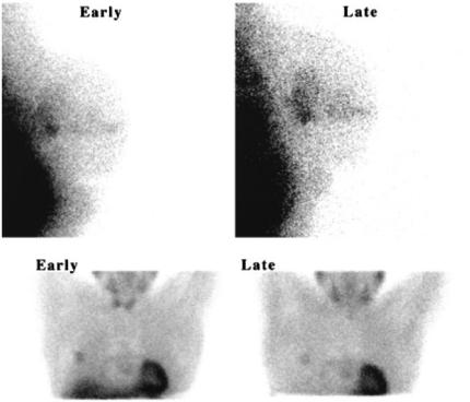
Primary intrinsic and/or acquired multidrug resistance (MDR) is the main obstacle to successful cancer treatment. Functional molecular imaging of MDR in cancer using single photon or positron emitters may be helpful to identify multidrug-resistant tumours and predict not only those patients who are resistant to treatment, with a clinically unfavourable prognosis, but also those who are susceptible to the development of drug toxicity or even certain tumours . Variations in the mdr1 gene product may directly affect the therapeutic effectiveness, and single nucleotide polymorphisms for the mdr1 gene may be associated with altered oral bioavailability of MDR1 substrates, drug resistance, and a susceptibility to some human diseases. The challenge of translating the concept of MDR modulation in vivo involves a complex cellular interplay between both malignant and normal cells. Integration and correlation of functional single photon emission tomography or positron emission tomography imaging findings with mdr1 genotype and clinical data may contribute to efficient management by selecting cancer patients with the appropriate molecular phenotype for maximal individual therapeutic benefit, as well as those who are non-responders. This review describes a role for functional imaging of classical mechanisms of MDR with an emphasis on readily available [(99m)Tc]MIBI scintigraphy. MIBI scintigraphy has been shown to be a non-invasive cost-effective in vivo assay of ATP-binding cassette transporters associated with MDR in cancer, including P-glycoprotein, multidrug-resistant protein 1 and breast cancer resistant protein. New imaging agents for molecular targets such as vascular endothelial growth factor and HER2 receptors, may potentially be combined with MDR imaging substrates to more accurately predict the therapeutic response to anticancer drugs, guiding individualised treatment while minimising the economic health costs of ineffective therapy in an era of personalised medicine.
原发性内在和/或获得性多药耐药(MDR)是癌症治疗成功的主要障碍。使用单光子或正电子发射体对癌症中的 MDR 进行功能分子成像,可能有助于识别多药耐药肿瘤,并不仅预测那些对治疗有抵抗力、临床预后不佳的患者,还预测那些容易发生药物毒性甚至某些肿瘤的患者。mdr1 基因产物的变异可能直接影响治疗效果,mdr1 基因的单核苷酸多态性可能与 MDR1 底物的口服生物利用度改变、耐药性以及某些人类疾病的易感性有关。将 MDR 调节的概念转化为体内的挑战涉及到恶性和正常细胞之间复杂的细胞相互作用。功能单光子发射断层扫描或正电子发射断层扫描成像结果与 mdr1 基因型和临床数据的整合和相关性,可能有助于通过选择具有适当分子表型的癌症患者,为最大化个体治疗获益以及非应答者提供有效的管理,从而选择对最大个体治疗获益具有适当分子表型的癌症患者。这篇综述描述了使用功能成像研究 MDR 经典机制的作用,重点是易于获得的 [(99m)Tc]MIBI 闪烁显像。MIBI 闪烁显像已被证明是一种非侵入性、具有成本效益的体内测定方法,可用于测定与癌症中的 MDR 相关的 ABC 转运蛋白,包括 P-糖蛋白、多药耐药蛋白 1 和乳腺癌耐药蛋白。新型分子靶点成像剂,如血管内皮生长因子和 HER2 受体,可能与 MDR 成像底物结合,更准确地预测对癌症药物的治疗反应,指导个体化治疗,同时在个体化医疗时代最小化无效治疗的经济健康成本。