University of Ottawa Heart Institute, Ottawa, ON K1Y 4W7, Canada.
Department of Biochemistry, Microbiology and Immunology, University of Ottawa, Ottawa, ON K1H 8M5, Canada.
Molecules. 2020 Dec 21;25(24):6042. doi: 10.3390/molecules25246042.
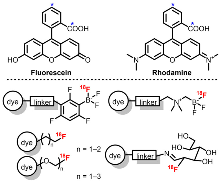
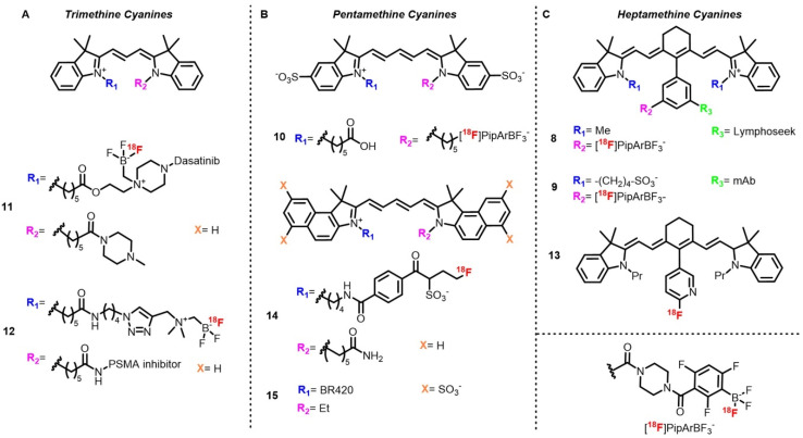
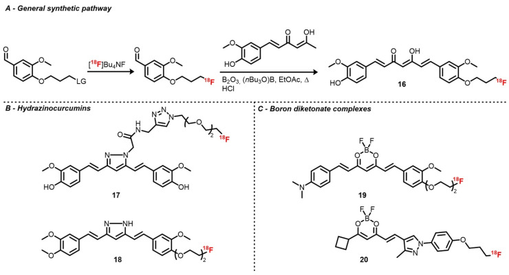
Recent progress realized in the development of optical imaging (OPI) probes and devices has made this technique more and more affordable for imaging studies and fluorescence-guided surgery procedures. However, this imaging modality still suffers from a low depth of penetration, thus limiting its use to shallow tissues or endoscopy-based procedures. In contrast, positron emission tomography (PET) presents a high depth of penetration and the resulting signal is less attenuated, allowing for imaging in-depth tissues. Thus, association of these imaging techniques has the potential to push back the limits of each single modality. Recently, several research groups have been involved in the development of radiolabeled fluorophores with the aim of affording dual-mode PET/OPI probes used in preclinical imaging studies of diverse pathological conditions such as cancer, Alzheimer's disease, or cardiovascular diseases. Among all the available PET-active radionuclides, F stands out as the most widely used for clinical imaging thanks to its advantageous characteristics (t = 109.77 min; 97% β emitter). This review focuses on the recent efforts in the synthesis and radiofluorination of fluorescent scaffolds such as 4,4-difluoro-4-bora-diazaindacenes (BODIPYs), cyanines, and xanthene derivatives and their use in preclinical imaging studies using both PET and OPI technologies.
近年来,光学成像(OPI)探针和设备的发展取得了重大进展,使得该技术在成像研究和荧光引导手术中越来越经济实惠。然而,这种成像方式仍然存在穿透深度低的问题,因此限制了其在浅层组织或内窥镜手术中的应用。相比之下,正电子发射断层扫描(PET)具有较高的穿透深度,得到的信号衰减较小,允许对深部组织进行成像。因此,这些成像技术的联合使用有可能突破每种单一模态的限制。最近,一些研究小组一直在致力于开发放射性标记荧光团,旨在提供用于临床前成像研究的双模式 PET/OPI 探针,用于研究各种病理情况,如癌症、阿尔茨海默病或心血管疾病。在所有可用的 PET 活性放射性核素中,F 因其优势特征(t = 109.77 分钟;97%β 发射器)而被广泛用于临床成像。本文重点介绍了近年来在合成和放射性标记荧光支架方面的努力,如 4,4-二氟-4-硼-二氮杂芴(BODIPYs)、花菁和呫吨衍生物,并将其用于使用 PET 和 OPI 技术的临床前成像研究。