INSERM U848, Villejuif F-94805, France.
Cell Death Dis. 2010;1(2):e25. doi: 10.1038/cddis.2010.6.
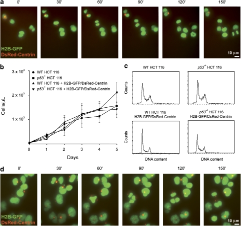
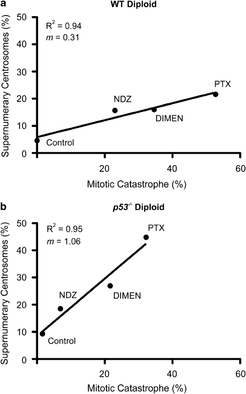
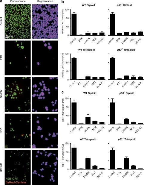
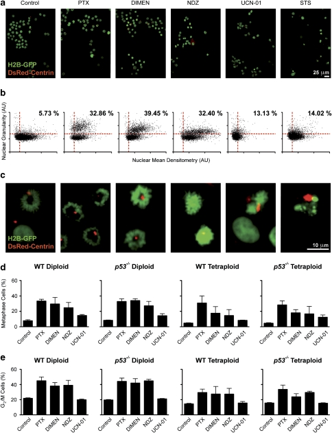
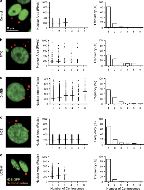
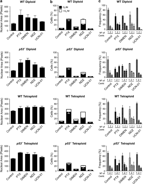
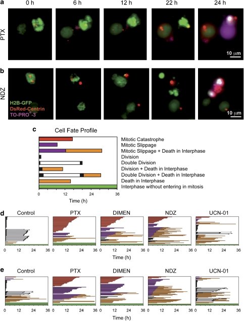
Mitotic catastrophe can be defined as a cell death mode that occurs during or shortly after a prolonged/aberrant mitosis, and can show apoptotic or necrotic features. However, conventional procedures for the detection of apoptosis or necrosis, including biochemical bulk assays and cytofluorometric techniques, cannot discriminate among pre-mitotic, mitotic and post-mitotic death, and hence are inappropriate to monitor mitotic catastrophe. To address this issue, we generated isogenic human colon carcinoma cell lines that differ in ploidy and p53 status, yet express similar amounts of fluorescent biosensors that allow for the visualization of chromatin (histone H2B coupled to green fluorescent protein (GFP)) and centrosomes (centrin coupled to the Discosoma striata red fluorescent protein (DsRed)). By combining high-resolution fluorescence videomicroscopy and automated image analysis, we established protocols and settings for the simultaneous assessment of ploidy, mitosis, centrosome number and cell death (which in our model system occurs mainly by apoptosis). Time-lapse videomicroscopy showed that this approach can be used for the high-throughput detection of mitotic catastrophe induced by three mechanistically distinct anti-mitotic agents (dimethylenastron (DIMEN), nocodazole (NDZ) and paclitaxel (PTX)), and - in this context - revealed an important role of p53 in the control of centrosome number.
有丝分裂灾难可被定义为一种在长时间/异常有丝分裂期间或之后发生的细胞死亡模式,并且可以显示凋亡或坏死特征。然而,用于检测凋亡或坏死的常规程序,包括生化批量测定和细胞荧光技术,不能区分有丝分裂前、有丝分裂中和有丝分裂后死亡,因此不适合监测有丝分裂灾难。为了解决这个问题,我们生成了具有不同倍性和 p53 状态的同基因人结肠癌细胞系,但表达相似量的荧光生物传感器,允许可视化染色质(与绿色荧光蛋白(GFP)偶联的组蛋白 H2B)和中心体(与 Discosoma striata 红色荧光蛋白(DsRed)偶联的中心粒)。通过结合高分辨率荧光视频显微镜和自动图像分析,我们建立了同时评估倍性、有丝分裂、中心体数量和细胞死亡(在我们的模型系统中主要通过凋亡发生)的方案和设置。延时视频显微镜显示,这种方法可用于高通量检测三种机制上不同的抗有丝分裂剂(二甲烯雄酮(DIMEN)、诺考达唑(NDZ)和紫杉醇(PTX))诱导的有丝分裂灾难,并且 - 在这种情况下 - 揭示了 p53 在控制中心体数量方面的重要作用。