Department of Physiology, School of Medicine, University of Pennsylvania, Philadelphia, Pennsylvania 19104-6085, USA.
J Cell Physiol. 2012 Jan;227(1):172-82. doi: 10.1002/jcp.22715.
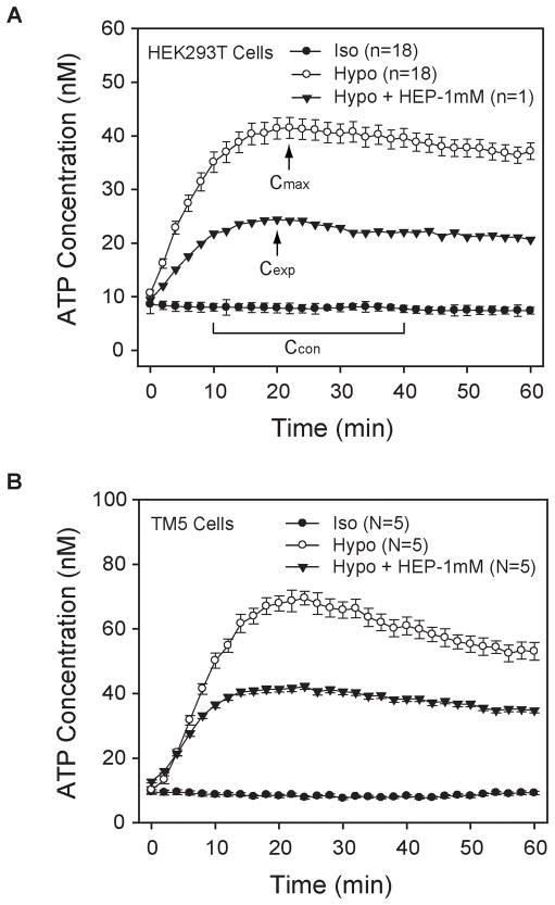
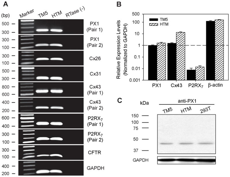
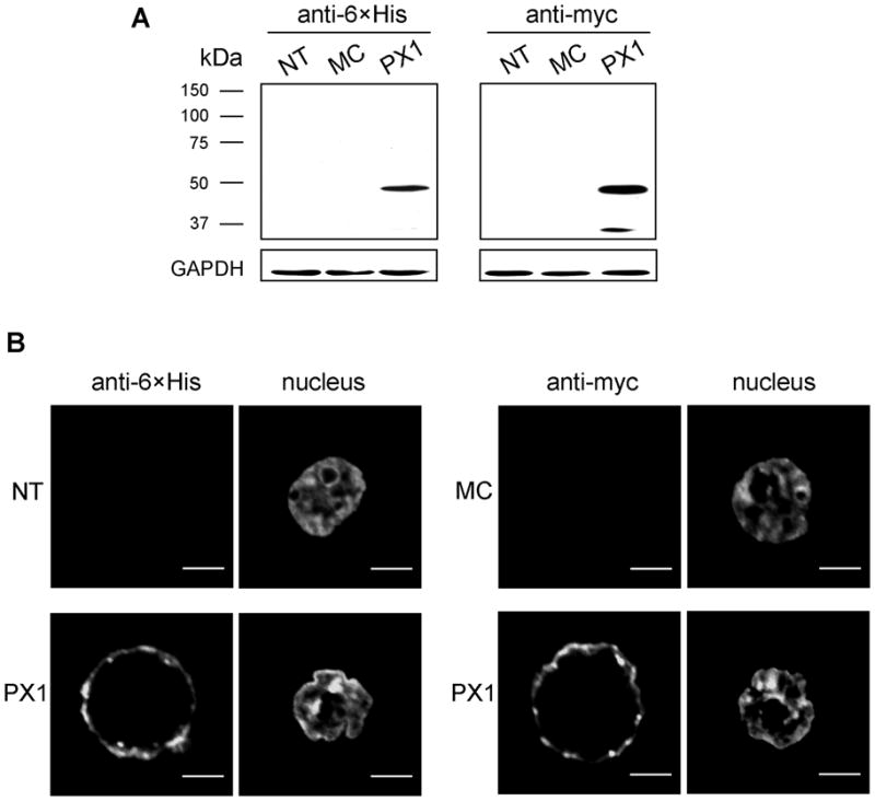
Our guiding hypothesis is that ecto-enzymatic conversion of extracellular ATP to adenosine activates A(1) adenosine receptors, reducing resistance to aqueous humor outflow and intraocular pressure. The initial step in this purinergic regulation is ATP release from outflow-pathway cells by mechanisms unknown. We measured similar ATP release from human explant-derived primary trabecular meshwork (TM) cells (HTM) and a human TM cell line (TM5). Responses to 21 inhibitors indicated that pannexin-1 (PX1) and connexin (Cx) hemichannels and P2X(7) receptors (P2RX(7) ) were comparably important in modulating ATP release induced by hypotonic swelling, whereas vesicular release was insignificant. Consistent with prior studies of PX1 activity in certain other cells, ATP release was lowered by the reducing agent dithiothreitol. Overexpressing PX1 in HEK293T cells promoted, while partial knockdown (KD) in both HEK293T and TM5 cells inhibited hypotonicity-activated ATP release. Additionally, KD reduced the pharmacologically defined contribution of PX1 and enhanced those of Cx and P2RX(7) . ATP release was also triggered by raising intracellular Ca(2+) activity with ionomycin after a prolonged lag time and was unaffected by the PX1 blocker probenecid, but nearly abolished by P2RX(7) antagonists. We conclude that swelling-stimulated ATP release from human TM cells is physiologically mediated by PX1 and Cx hemichannels and P2X(7) receptors, but not by vesicular release. PX1 appears not to be stimulated by intracellular Ca(2+) in TM cells, but can be modulated by oxidation-reduction state. The P2RX(7) -dependent component of swelling-activated release may be mediated by PX1 hemichannels or reflect apoptotic magnification of ATP release, either through itself and/or hemichannels.
我们的指导假说是,细胞外 ATP 向腺苷的外切酶转化激活 A(1) 腺苷受体,降低房水流出和眼内压的阻力。这种嘌呤能调节的初始步骤是未知机制从流出途径细胞释放 ATP。我们测量了人源性小梁网(TM)细胞(HTM)和人 TM 细胞系(TM5)的类似 ATP 释放。对 21 种抑制剂的反应表明,在调节由低渗肿胀诱导的 ATP 释放方面,连接蛋白(Cx)半通道和 P2X(7) 受体(P2RX(7) )与 Pannexin-1(PX1)同样重要,而囊泡释放则无足轻重。与先前在某些其他细胞中 PX1 活性的研究一致,还原剂二硫苏糖醇降低了 ATP 释放。在 HEK293T 细胞中过表达 PX1 促进,而在 HEK293T 和 TM5 细胞中部分敲低(KD)抑制低渗激活的 ATP 释放。此外,KD 降低了药理学上定义的 PX1 贡献,增强了 Cx 和 P2RX(7) 的贡献。用离子霉素长时间延迟后,升高细胞内 Ca(2+) 活性也会触发 ATP 释放,而 PX1 阻断剂丙磺舒对其无影响,但几乎完全消除了 P2RX(7) 拮抗剂的作用。我们的结论是,人 TM 细胞的肿胀刺激的 ATP 释放是由 PX1 和 Cx 半通道和 P2X(7) 受体介导的,而不是由囊泡释放介导的。在 TM 细胞中,PX1 似乎不受细胞内 Ca(2+) 的刺激,但可以通过氧化还原状态进行调节。肿胀激活释放的 P2RX(7) 依赖性成分可能通过 PX1 半通道介导,或者通过自身和/或半通道放大 ATP 释放来反映细胞凋亡。