Agarwal Amit, Bhake Arvind, Kakani Anand
Department of Neurosurgery, Datta Meghe Institute of Medical Sciences, Wardha, Maharashtra, India.
Asian Spine J. 2011 Mar;5(1):59-63. doi: 10.4184/asj.2011.5.1.59. Epub 2011 Mar 2.
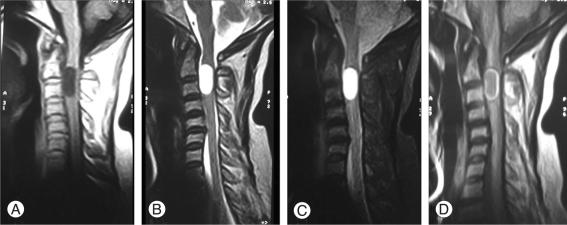
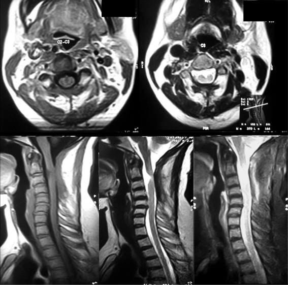
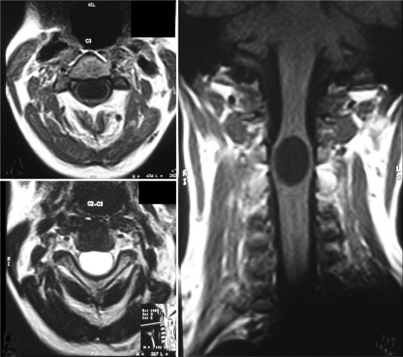
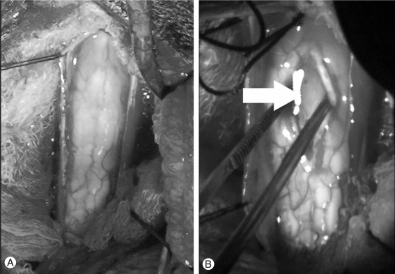
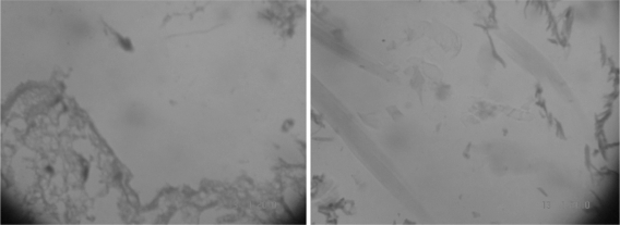
Intramedullary spinal epidermoid cysts are benign ectopic embryological growths with reported incidence of less than 1% of intramedullary tumors. In this case we report an unusual cervical intramedullary epidermid with liquid contents. A 40-year-old patient presented with progressive weakness of all four limbs of four months duration, bowel and bladder disturbances of two days duration, pain and paresthesias in all four limbs. Magnetic resonance imaging (MRI) revealed a well defined intramedullary lesion extending from C2-C3 level with widening of the cord. The lesion was hypointense on T1W images, hyperintense on T2W and fluid attenuation and inversion recovery images with thin rim of enhancement after contrast administration. Histopathological examination of the excised specimen revealed epidermal lining and keratinous material features of an epidermoid cyst. As in present case, rarely epidermoid cyst can have clear contents, and an MRI finding can closely mimic the features of arachnoid cyst, findings not classical and is different than described in literature.
脊髓髓内表皮样囊肿是良性异位胚胎发育性病变,据报道其发病率在髓内肿瘤中不到1%。在本病例中,我们报告了一例罕见的颈椎髓内表皮样囊肿,其内容物为液体。一名40岁患者出现四肢进行性无力4个月,伴有2天的肠道和膀胱功能障碍,以及四肢疼痛和感觉异常。磁共振成像(MRI)显示一个边界清晰的髓内病变,从C2 - C3水平延伸,脊髓增宽。该病变在T1加权图像上呈低信号,在T2加权图像和液体衰减反转恢复图像上呈高信号,增强后有薄边缘强化。切除标本的组织病理学检查显示为表皮样囊肿的表皮内衬和角质物质特征。如本病例所示,表皮样囊肿很少会有清亮内容物,其MRI表现可与蛛网膜囊肿的特征极为相似,这些表现并不典型,与文献报道不同。