Division of Rheumatology, Research Institute of the McGill University Health Centre, McGill University, Montreal, Quebec H3G 1A4, Canada.
J Immunol. 2011 Apr 15;186(8):4771-81. doi: 10.4049/jimmunol.1000921. Epub 2011 Mar 9.
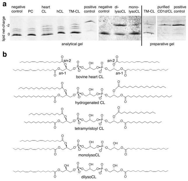
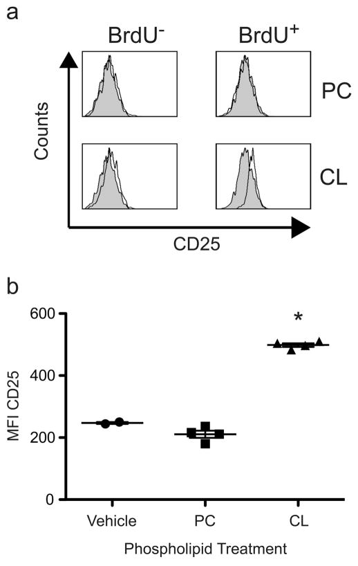
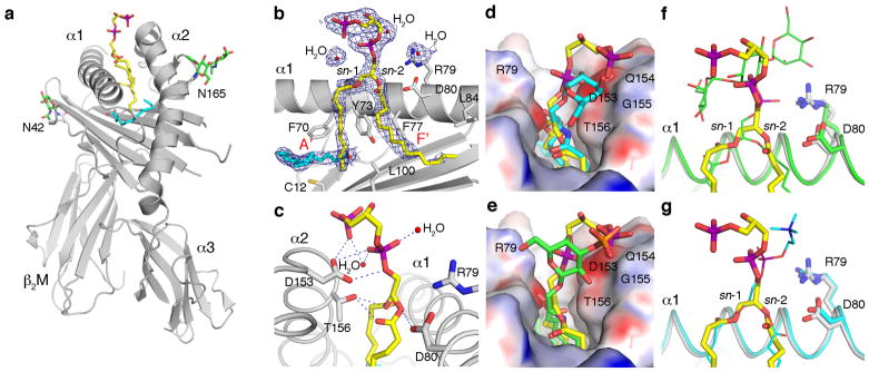
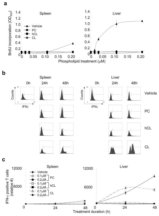
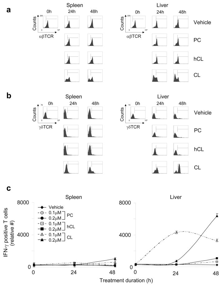
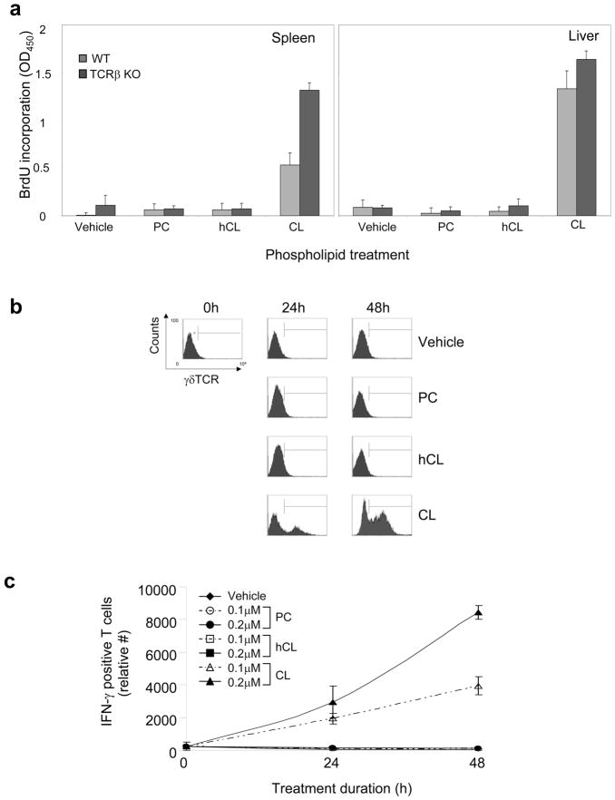
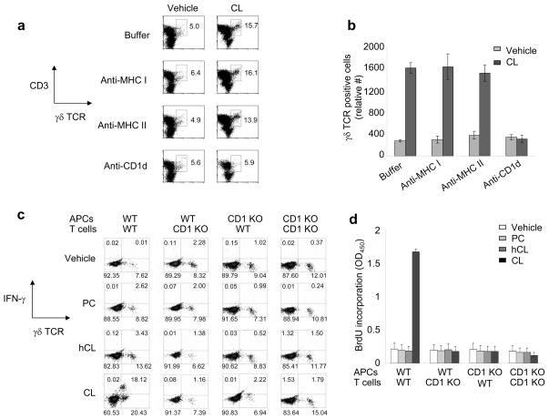
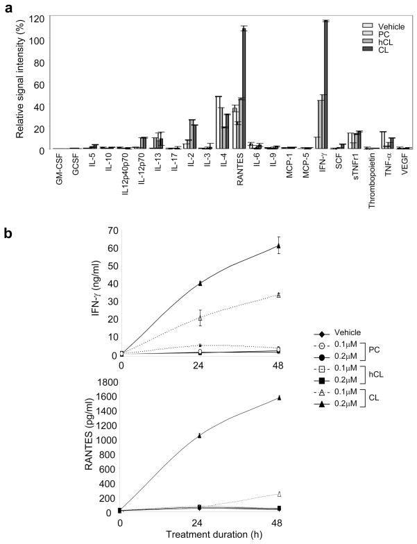
Cardiolipin (CL), a major phospholipid in bacterial cell walls, is sequestered from the immune system in mammalian mitochondria and is, therefore, a potential danger signal. Based on growing evidence that phospholipids constitute natural ligands for CD1 and that CD1d-restricted T cells recognize phospholipids, we hypothesized that CD1d binds and presents CL and that T cells in the normal immune repertoire respond to CL in a CD1d-restricted manner. We determined the murine CD1d-CL crystal structure at 2.3 Å resolution and established through additional lipid loading experiments that CL, a tetra-acylated phospholipid, binds to murine CD1d with two alkyl chains buried inside the CD1d binding groove and the remaining two exposed into the solvent. We furthermore demonstrate the functional stimulatory activity of CL, showing that splenic and hepatic γδ T cells from healthy mice proliferate in vitro in response to mammalian or bacterial CL in a dose-dependent and CD1d-restricted manner, rapidly secreting the cytokines IFN-γ and RANTES. Finally, we show that hepatic γδ T cells are activated in vivo by CD1d-bearing dendritic cells that have been pulsed with CL, but not phosphatidylcholine. Together, these findings demonstrate that CD1d is able to bind and present CL to a subset of CL-responsive γδ T cells that exist in the spleen and liver of healthy mice and suggest that these cells could play a role in host responses to bacterial lipids and, potentially, self-CL. We propose that CL-responsive γδ T cells play a role in immune surveillance during infection and tissue injury.
心磷脂(CL)是细菌细胞壁中的一种主要磷脂,在哺乳动物的线粒体中与免疫系统隔离,因此是一种潜在的危险信号。基于越来越多的证据表明磷脂是 CD1 的天然配体,并且 CD1d 限制性 T 细胞识别磷脂,我们假设 CD1d 结合并呈递 CL,并且正常免疫谱中的 T 细胞以 CD1d 限制性方式对 CL 产生反应。我们确定了 2.3Å 分辨率的鼠 CD1d-CL 晶体结构,并通过额外的脂质加载实验证实,CL 是一种四酰化磷脂,其两条烷基链埋藏在 CD1d 结合槽内,其余两条暴露在溶剂中,与鼠 CD1d 结合。我们还证明了 CL 的功能刺激活性,表明来自健康小鼠的脾和肝γδ T 细胞在体外以剂量依赖和 CD1d 限制的方式对哺乳动物或细菌 CL 增殖,并迅速分泌细胞因子 IFN-γ 和 RANTES。最后,我们表明,通过用 CL 脉冲的携带 CD1d 的树突状细胞,体内激活肝γδ T 细胞,但不能激活磷酸胆碱。总之,这些发现表明 CD1d 能够结合并呈递 CL 给存在于健康小鼠脾脏和肝脏中的一组 CL 反应性 γδ T 细胞,并表明这些细胞可能在宿主对细菌脂质的反应中发挥作用,并且可能在自身 CL 中发挥作用。我们提出,CL 反应性 γδ T 细胞在感染和组织损伤期间的免疫监视中发挥作用。