de Duve Institute, Université Catholique de Louvain, Brussels, Belgium.
Hepatology. 2011 Jun;53(6):1959-66. doi: 10.1002/hep.24292. Epub 2011 May 2.
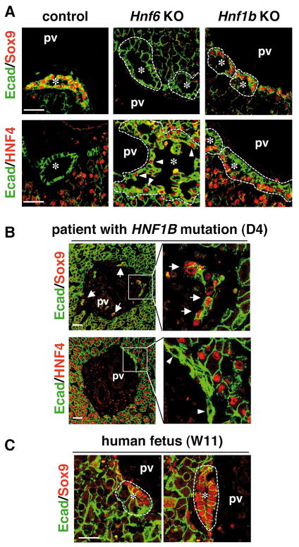
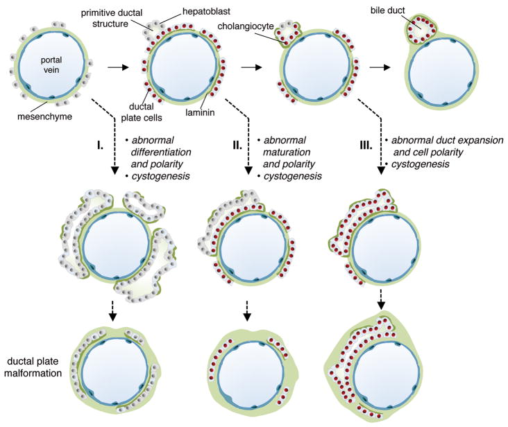
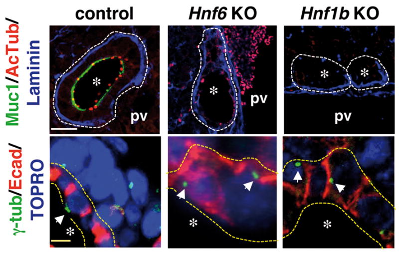
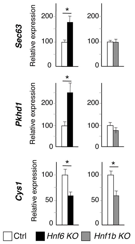
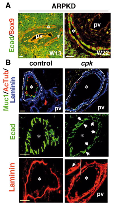
Ductal plate malformations (DPMs) are developmental anomalies considered to result from lack of ductal plate remodeling during bile duct morphogenesis. In mice, bile duct development is initiated by the formation of primitive ductal structures lined by two cell types, namely ductal plate cells and hepatoblasts. During ductal plate remodeling, the primitive ductal structures mature to ducts as a result from differentiation of the ductal plate cells and hepatoblasts to cholangiocytes. Here, we report this process is conserved in human fetal liver. These findings prompted us to evaluate how DPMs develop in three mouse models, namely mice with livers deficient in hepatocyte nuclear factor 6 (HNF6), HNF1β, or cystin-1 (cpk [congenital polycystic kidney] mice). Human liver from a patient with a HNF1B/TCF2 mutation, and from fetuses affected with autosomal recessive polycystic kidney disease (ARPKD) were also analyzed. Despite the epistatic relationship between HNF6, HNF1β, and cystin-1, the three mouse models displayed distinct morphogenic mechanisms of DPM. They all developed biliary cysts lined by cells with abnormal apicobasal polarity. However, the absence of HNF6 led to an early defect in ductal plate cell differentiation. In HNF1β-deficient liver, maturation of the primitive ductal structures was impaired. Normal differentiation and maturation but abnormal duct expansion was apparent in cpk mouse livers and in human fetal ARPKD.
DPM is the common endpoint of distinct defects initiated at distinct stages of bile duct morphogenesis. Our observations provide a new pathogenic classification of DPM.
管板畸形(DPM)被认为是由于胆管形态发生过程中缺乏管板重塑而导致的发育异常。在小鼠中,胆管发育是由两种细胞类型(即胆管板细胞和肝母细胞)衬里的原始胆管结构的形成开始的。在胆管板重塑过程中,原始胆管结构由于胆管板细胞和肝母细胞向胆管细胞分化而成熟为胆管。在这里,我们报告在人类胎儿肝脏中发现了这一过程。这些发现促使我们评估 DPM 如何在三种小鼠模型中发育,即肝脏缺乏肝细胞核因子 6(HNF6)、HNF1β或胱氨酸-1(cpk [先天性多囊肾病]小鼠)的小鼠、患有 HNF1B/TCF2 突变的患者的人类肝脏,以及受常染色体隐性多囊肾病(ARPKD)影响的胎儿的肝脏。尽管 HNF6、HNF1β和胱氨酸-1之间存在上位关系,但三种小鼠模型显示出不同的 DPM 形态发生机制。它们都形成了由具有异常顶底极性的细胞衬里的胆管囊肿。然而,HNF6 的缺失导致胆管板细胞分化的早期缺陷。在 HNF1β 缺陷的肝脏中,原始胆管结构的成熟受到损害。在 cpk 小鼠肝脏和人类胎儿 ARPKD 中,观察到正常的分化和成熟,但异常的胆管扩张。
DPM 是在胆管形态发生的不同阶段开始的不同缺陷的共同终点。我们的观察结果为 DPM 提供了一种新的致病分类。