• 文献检索
  • 文档翻译
  • 深度研究
  • 学术资讯
  • Suppr Zotero 插件Zotero 插件
  • 邀请有礼
  • 套餐&价格
  • 历史记录
应用&插件
Suppr Zotero 插件Zotero 插件浏览器插件Mac 客户端Windows 客户端微信小程序
定价
高级版会员购买积分包购买API积分包
服务
文献检索文档翻译深度研究API 文档MCP 服务
关于我们
关于 Suppr公司介绍联系我们用户协议隐私条款
关注我们

Suppr 超能文献

核心技术专利:CN118964589B侵权必究
粤ICP备2023148730 号-1Suppr @ 2026

文献检索

告别复杂PubMed语法,用中文像聊天一样搜索,搜遍4000万医学文献。AI智能推荐,让科研检索更轻松。

立即免费搜索

文件翻译

保留排版,准确专业,支持PDF/Word/PPT等文件格式,支持 12+语言互译。

免费翻译文档

深度研究

AI帮你快速写综述,25分钟生成高质量综述,智能提取关键信息,辅助科研写作。

立即免费体验

功能磁共振:乳腺癌反应的生物标志物。

Functional magnetic resonance: biomarkers of response in breast cancer.

机构信息

Clinical Magnetic Resonance Group, Institute of Cancer Research, Royal Marsden NHS Foundation Trust, Sutton, Surrey, SM2 5PT, UK.

出版信息

Breast Cancer Res. 2011 Feb 23;13(1):204. doi: 10.1186/bcr2815.

DOI:10.1186/bcr2815
PMID:21392409
原文链接:https://pmc.ncbi.nlm.nih.gov/articles/PMC3109577/
Abstract

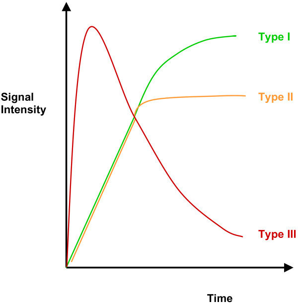
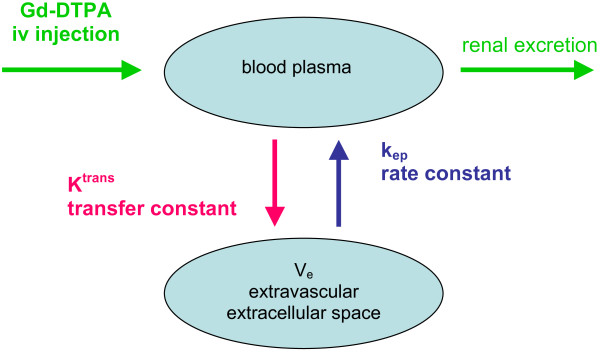
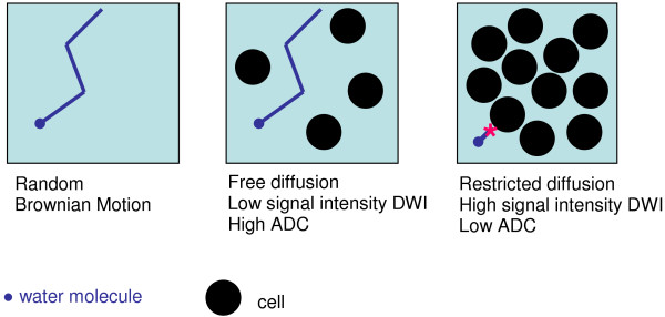
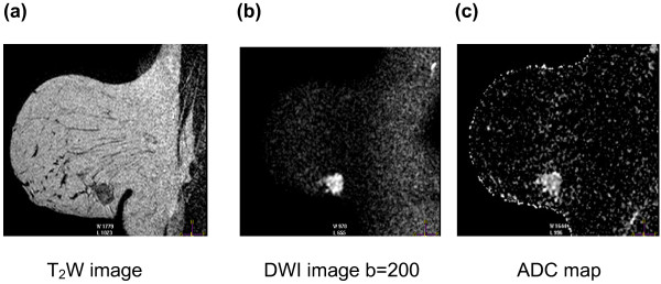
Functional magnetic resonance (MR) encompasses a spectrum of techniques that depict physiological and molecular processes before morphological changes are visible on conventional imaging. As understanding of the pathophysiological and biomolecular processes involved in breast malignancies evolves, newer functional MR techniques can be employed that define early predictive and surrogate biomarkers for monitoring response to chemotherapy. Neoadjuvant chemotherapy is increasingly used in women with primary breast malignancies to down-stage the tumour and enable successful breast conservation surgery. It also plays a role in the treatment of undetected micrometastases. Cardinal physiological features of tumours that occur as a result of interactions between cancer cells, stromal cells and secreted factors and cytokines and how they change with treatment provide the opportunity to detect changes in the tumour microenvironment prior to any morphological change. Through sequential imaging, tumour response can be assessed and non-responders can be identified early to enable alternative therapies to be considered. This review summarises the functional magnetic resonance biomarkers of response in patients with breast cancer that are currently available and under development. We describe the current state of each biomarker and explore their potential clinical uses and limitations in assessing treatment response. With the aid of selected interesting cases, biomarkers related to dynamic contrast-enhanced MRI, diffusion-weighted MRI, T2*/BOLD and MR spectroscopy are described and illustrated. The potential of newer approaches, such as MR elastography, are also reviewed.

摘要

功能磁共振(MR)涵盖了一系列技术,可在常规成像可见形态变化之前描绘生理和分子过程。随着对乳腺癌相关病理生理和生物分子过程的认识不断发展,可采用新的功能磁共振技术来定义早期预测和替代生物标志物,以监测化疗反应。新辅助化疗越来越多地用于原发性乳腺癌女性,以降低肿瘤分期并实现成功的保乳手术。它在治疗未检测到的微转移中也发挥作用。由于癌细胞、基质细胞和分泌因子和细胞因子之间的相互作用而发生的肿瘤的主要生理特征,以及它们如何随治疗而变化,为在任何形态变化之前检测肿瘤微环境的变化提供了机会。通过连续成像,可以评估肿瘤反应,早期识别无反应者,从而考虑替代治疗方法。这篇综述总结了目前可用于和正在开发的乳腺癌患者功能磁共振反应的生物标志物。我们描述了每个生物标志物的现状,并探讨了它们在评估治疗反应中的潜在临床应用和局限性。通过选择一些有趣的病例,描述并说明了与动态对比增强 MRI、弥散加权 MRI、T2*/BOLD 和磁共振波谱相关的生物标志物。还回顾了新方法(如磁共振弹性成像)的潜力。

https://cdn.ncbi.nlm.nih.gov/pmc/blobs/acbf/3109577/c26dfe1777fd/bcr2815-6.jpg
https://cdn.ncbi.nlm.nih.gov/pmc/blobs/acbf/3109577/99f7e6bec156/bcr2815-1.jpg
https://cdn.ncbi.nlm.nih.gov/pmc/blobs/acbf/3109577/918af77f78aa/bcr2815-2.jpg
https://cdn.ncbi.nlm.nih.gov/pmc/blobs/acbf/3109577/0902a52c8f2d/bcr2815-3.jpg
https://cdn.ncbi.nlm.nih.gov/pmc/blobs/acbf/3109577/b1a92ff09cc2/bcr2815-4.jpg
https://cdn.ncbi.nlm.nih.gov/pmc/blobs/acbf/3109577/89ae64c0c5af/bcr2815-5.jpg
https://cdn.ncbi.nlm.nih.gov/pmc/blobs/acbf/3109577/c26dfe1777fd/bcr2815-6.jpg

相似文献

1
Functional magnetic resonance: biomarkers of response in breast cancer.功能磁共振:乳腺癌反应的生物标志物。
Breast Cancer Res. 2011 Feb 23;13(1):204. doi: 10.1186/bcr2815.
2
[Molecular breast imaging. An update].[分子乳腺成像。最新进展]
Radiologe. 2014 Mar;54(3):241-53. doi: 10.1007/s00117-013-2580-8.
3
Multiparametric MRI With Dynamic Contrast Enhancement, Diffusion-Weighted Imaging, and 31-Phosphorus Spectroscopy at 7 T for Characterization of Breast Cancer.7T下采用动态对比增强、扩散加权成像和31磷波谱的多参数MRI用于乳腺癌特征分析
Invest Radiol. 2015 Nov;50(11):766-71. doi: 10.1097/RLI.0000000000000183.
4
Multiparametric MR Imaging of Breast Cancer.乳腺癌的多参数磁共振成像
Magn Reson Imaging Clin N Am. 2016 Feb;24(1):223-238. doi: 10.1016/j.mric.2015.08.012.
5
Multiparametric MR Imaging in Abdominal Malignancies.腹部恶性肿瘤的多参数磁共振成像
Magn Reson Imaging Clin N Am. 2016 Feb;24(1):157-186. doi: 10.1016/j.mric.2015.08.005.
6
Investigating the contribution of hyaluronan to the breast tumour microenvironment using multiparametric MRI and MR elastography.使用多参数 MRI 和 MR 弹性成像研究透明质酸对乳腺肿瘤微环境的贡献。
Mol Oncol. 2023 Jun;17(6):1076-1092. doi: 10.1002/1878-0261.13437. Epub 2023 May 3.
7
Assessment of Hypoxia in Breast Cancer: Emerging Functional MR Imaging and Spectroscopy Techniques and Clinical Applications.乳腺癌中缺氧的评估:新兴的功能磁共振成像和光谱技术及临床应用
J Magn Reson Imaging. 2025 Jan;61(1):83-96. doi: 10.1002/jmri.29424. Epub 2024 May 4.
8
Utilisation of MR spectroscopy and diffusion weighted imaging in predicting and monitoring of breast cancer response to chemotherapy.磁共振波谱成像和扩散加权成像在预测和监测乳腺癌化疗反应中的应用
J Med Imaging Radiat Oncol. 2015 Jun;59(3):268-77. doi: 10.1111/1754-9485.12310. Epub 2015 Apr 24.
9
Assessing response to treatment in breast cancer using magnetic resonance.利用磁共振成像评估乳腺癌的治疗反应。
J Exp Clin Cancer Res. 2002 Sep;21(3 Suppl):39-45.
10
Identification of residual breast carcinoma following neoadjuvant chemotherapy: diffusion-weighted imaging--comparison with contrast-enhanced MR imaging and pathologic findings.新辅助化疗后残余乳腺癌的识别:弥散加权成像——与对比增强磁共振成像和病理发现的比较。
Radiology. 2010 Feb;254(2):357-66. doi: 10.1148/radiol.2542090405.

引用本文的文献

1
Hyperoxic BOLD-MRI-Based Characterization of Breast Cancer Molecular Subtypes Is Independent of the Supplied Amount of Oxygen: A Preclinical Study.基于高氧血氧水平依赖性功能磁共振成像对乳腺癌分子亚型的特征描述与氧气供应量无关:一项临床前研究
Diagnostics (Basel). 2023 Sep 14;13(18):2946. doi: 10.3390/diagnostics13182946.
2
Functional Imaging of Hypoxia: PET and MRI.缺氧的功能成像:正电子发射断层扫描(PET)和磁共振成像(MRI)
Cancers (Basel). 2023 Jun 25;15(13):3336. doi: 10.3390/cancers15133336.
3
Hyperpolarized Carbon-13 MRI in Breast Cancer.乳腺癌中的超极化碳-13磁共振成像

本文引用的文献

1
Multiparametric magnetic resonance imaging, spectroscopy and multinuclear (²³Na) imaging monitoring of preoperative chemotherapy for locally advanced breast cancer.多参数磁共振成像、波谱和多核(²³Na)成像监测局部晚期乳腺癌术前化疗。
Acad Radiol. 2010 Dec;17(12):1477-85. doi: 10.1016/j.acra.2010.07.009. Epub 2010 Sep 21.
2
Primary human breast adenocarcinoma: imaging and histologic correlates of intrinsic susceptibility-weighted MR imaging before and during chemotherapy.原发性人乳腺腺癌:化疗前后磁共振成像固有敏感性加权成像的影像学和组织学相关性。
Radiology. 2010 Dec;257(3):643-52. doi: 10.1148/radiol.10100421. Epub 2010 Sep 21.
3
Diagnostics (Basel). 2023 Jul 7;13(13):2311. doi: 10.3390/diagnostics13132311.
4
Head-to-head comparison of perfluorobutane contrast-enhanced US and multiparametric MRI for breast cancer: a prospective, multicenter study.对比氟丙烷对比增强超声与多参数 MRI 用于乳腺癌的头对头研究:一项前瞻性、多中心研究。
Breast Cancer Res. 2023 May 30;25(1):61. doi: 10.1186/s13058-023-01650-3.
5
Multitasking dynamic contrast enhanced magnetic resonance imaging can accurately differentiate chronic pancreatitis from pancreatic ductal adenocarcinoma.多任务动态对比增强磁共振成像能够准确区分慢性胰腺炎和胰腺导管腺癌。
Front Oncol. 2023 Jan 6;12:1007134. doi: 10.3389/fonc.2022.1007134. eCollection 2022.
6
Overview of Artificial Intelligence in Breast Cancer Medical Imaging.乳腺癌医学成像中的人工智能概述
J Clin Med. 2023 Jan 4;12(2):419. doi: 10.3390/jcm12020419.
7
Dynamic contrast enhanced MRI of pulmonary adenocarcinomas for early risk stratification: higher contrast uptake associated with response and better prognosis.动态对比增强 MRI 对肺腺癌的早期风险分层:较高的对比摄取与反应和更好的预后相关。
BMC Med Imaging. 2022 Dec 5;22(1):215. doi: 10.1186/s12880-022-00943-x.
8
MRI as a biomarker for breast cancer diagnosis and prognosis.磁共振成像作为乳腺癌诊断和预后的生物标志物
BJR Open. 2022 May 26;4(1):20220002. doi: 10.1259/bjro.20220002. eCollection 2022.
9
Association of breast cancer risk, density, and stiffness: global tissue stiffness on breast MR elastography (MRE).乳腺癌风险、密度和硬度的关联:乳腺磁共振弹性成像(MRE)的全球组织硬度。
Breast Cancer Res Treat. 2022 Jul;194(1):79-89. doi: 10.1007/s10549-022-06607-2. Epub 2022 Apr 30.
10
Dynamic Contrast-enhanced and Diffusion-weighted Magnetic Resonance Imaging for Response Evaluation After Single-Dose Ablative Neoadjuvant Partial Breast Irradiation.动态对比增强和扩散加权磁共振成像用于单次消融新辅助部分乳腺照射后反应评估
Adv Radiat Oncol. 2021 Nov 20;7(2):100854. doi: 10.1016/j.adro.2021.100854. eCollection 2022 Mar-Apr.
Diffusion-weighted imaging improves the diagnostic accuracy of conventional 3.0-T breast MR imaging.
弥散加权成像提高了常规 3.0-T 乳腺磁共振成像的诊断准确性。
Radiology. 2010 Jul;256(1):64-73. doi: 10.1148/radiol.10091367.
4
Diffusion-weighted magnetic resonance imaging for pretreatment prediction and monitoring of treatment response of patients with locally advanced breast cancer undergoing neoadjuvant chemotherapy.弥散加权磁共振成像在局部进展期乳腺癌新辅助化疗患者治疗前预测和疗效监测中的应用。
Acta Oncol. 2010 Apr;49(3):354-60. doi: 10.3109/02841861003610184.
5
Predicting pathological response to neoadjuvant chemotherapy in breast cancer with quantitative 1H MR spectroscopy using the external standard method.采用外标法的 1H MR 波谱定量分析预测乳腺癌新辅助化疗的病理反应。
J Magn Reson Imaging. 2010 Apr;31(4):895-902. doi: 10.1002/jmri.22118.
6
Magnetic resonance imaging of the breast: recommendations from the EUSOMA working group.乳房磁共振成像:EUSOMA 工作组的建议。
Eur J Cancer. 2010 May;46(8):1296-316. doi: 10.1016/j.ejca.2010.02.015. Epub 2010 Mar 19.
7
Differential diagnosis of mammographically and clinically occult breast lesions on diffusion-weighted MRI.弥散加权 MRI 对乳腺触诊和 X 线钼靶摄影阴性的乳腺病变的鉴别诊断。
J Magn Reson Imaging. 2010 Mar;31(3):562-70. doi: 10.1002/jmri.22078.
8
Preliminary study of early response to neoadjuvant chemotherapy after the first cycle in breast cancer: comparison of 1H magnetic resonance spectroscopy with diffusion magnetic resonance imaging.乳腺癌新辅助化疗第一周期后早期疗效的初步研究:1H 磁共振波谱与弥散磁共振成像的比较。
Jpn J Radiol. 2010 Feb;28(2):101-9. doi: 10.1007/s11604-009-0391-7. Epub 2010 Feb 26.
9
B1 transmission-field inhomogeneity and enhancement ratio errors in dynamic contrast-enhanced MRI (DCE-MRI) of the breast at 3T.3T 下乳腺动态对比增强磁共振成像(DCE-MRI)中的 B1 传输场不均匀性和增强率误差
J Magn Reson Imaging. 2010 Jan;31(1):234-9. doi: 10.1002/jmri.22018.
10
Quantitative diffusion-weighted imaging as an adjunct to conventional breast MRI for improved positive predictive value.定量扩散加权成像作为常规乳腺 MRI 的辅助手段,可提高阳性预测值。
AJR Am J Roentgenol. 2009 Dec;193(6):1716-22. doi: 10.2214/AJR.08.2139.