Medical College of Wisconsin, Milwaukee, WI 53226, USA.
Arterioscler Thromb Vasc Biol. 2011 Jun;31(6):1351-6. doi: 10.1161/ATVBAHA.111.225334. Epub 2011 Mar 10.
Experimental studies of large-vessel thrombosis have been adapted for applications in mice, but they proffer limited quantifiable information in outcome measures. This study presents a novel approach for evaluating large-vessel thrombogenesis with temporally/spatially quantifiable measures and normalization methods for interanimal comparisons.
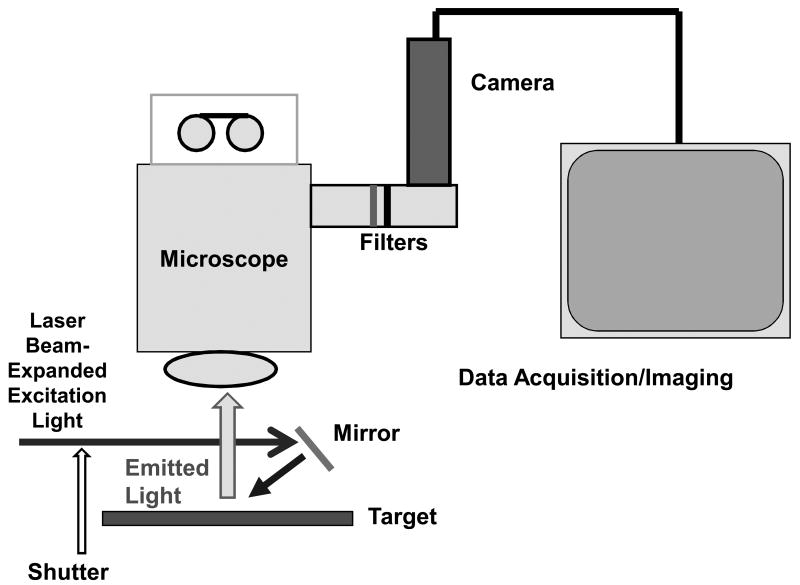
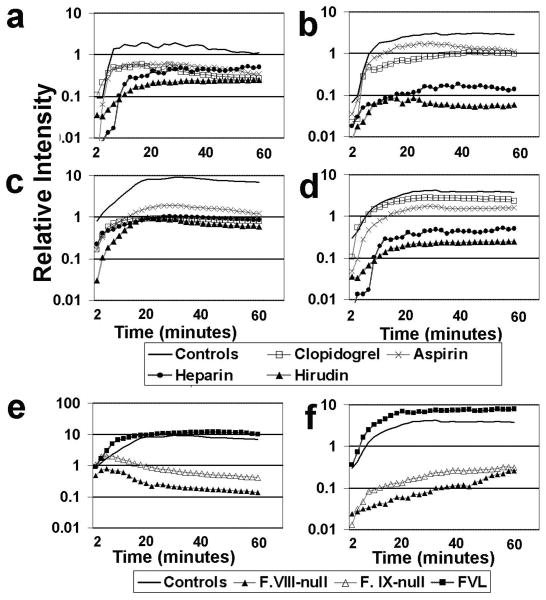
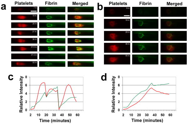
Shuttered, beam-expanded lasers provided uniform narrow-wavelength illumination of a ×100 microsurgical field with a large depth of focus. Thrombosis was generated in murine carotid arteries and femoral veins by brief vascular surface electrolytic injury. Thrombus-targeting fluorophores were injected systemically and subsequently localized at the site of thrombus induction. A low-light digital video camera with filter wheel provided target-specific image acquisition over a 60-minute interval. Platelets accumulated with a subsequent fibrin border emerging to stabilize the clot in both arteries and veins. Coagulation enzyme complexes colocalized with fibrin deposition. Large arteries underwent cyclic massive thromboembolization, whereas veins showed gradual shedding of microemboli and clot contraction. Systemic administration of fibrin- and platelet-inhibiting compounds reduced their respective targets but also often inhibited their clotting counterparts (platelets and fibrin, respectively) in both arteries and veins.
Intermediate-level magnified image capture represents a novel approach for analysis of fluorescence-based in vivo imaging, with quantitative application to the study of large-vessel thrombosis.
大血管血栓形成的实验研究已经适应于在小鼠中的应用,但它们在结果测量中提供的可量化信息有限。本研究提出了一种新的方法,用于评估大血管血栓形成,并提供时间/空间可量化的测量和用于动物间比较的归一化方法。
带快门的扩展光束激光器为 ×100 手术显微镜视野提供了均匀的窄波长照明,具有很大的景深。通过短暂的血管表面电解损伤在小鼠颈动脉和股静脉中产生血栓。血栓靶向荧光染料系统注射,并随后在血栓诱导部位定位。带有滤光轮的低光数字摄像机提供了在 60 分钟间隔内的目标特异性图像采集。血小板聚集,随后出现纤维蛋白边界,稳定了动脉和静脉中的血栓。凝血酶复合物与纤维蛋白沉积共定位。大动脉经历周期性的大量血栓栓塞,而静脉则逐渐脱落微栓子并收缩血栓。纤维蛋白和血小板抑制化合物的全身给药减少了它们各自的靶标,但也经常抑制了动脉和静脉中它们的凝血对应物(血小板和纤维蛋白)。
中级放大图像捕获代表了一种新的分析荧光体内成像的方法,可定量应用于大血管血栓形成的研究。