Department of Pediatrics, David Geffen School of Medicine at UCLA, A2-383 MDCC, 650 Charles Young Drive, Los Angeles, CA 90095, USA.
Pediatr Nephrol. 2011 Dec;26(12):2143-51. doi: 10.1007/s00467-011-1829-6. Epub 2011 Mar 11.
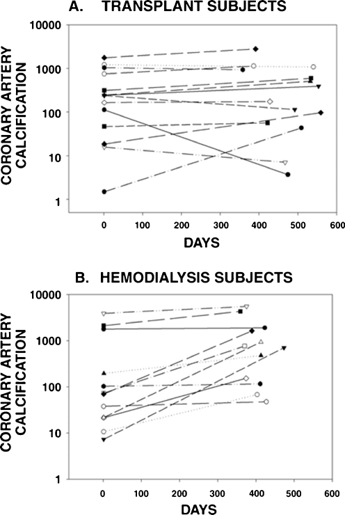
Successful kidney transplantation corrects many of the metabolic abnormalities associated with chronic kidney disease (CKD); however, skeletal and cardiovascular morbidity remain prevalent in pediatric kidney transplant recipients and current recommendations from the Kidney Disease Improving Global Outcomes (KDIGO) working group suggest that bone disease-including turnover, mineralization, volume, linear growth, and strength-as well as cardiovascular disease be evaluated in all patients with CKD. Although few studies have examined bone histology after renal transplantation, current data suggest that bone turnover and mineralization are altered in the majority of patients and that biochemical parameters are poor predictors of bone histology in this population. Dual energy X-ray absorptiometry (DXA) scanning, although widely performed, has significant limitations in the pediatric transplant population and values have not been shown to correlate with fracture risk; thus, DXA is not recommended as a tool for the assessment of bone density. Newer imaging techniques, including computed tomography (quantitative CT (QCT), peripheral QCT (pQCT), high resolution pQCT (HR-pQCT) and magnetic resonance imaging (MRI)), which provide volumetric assessments of bone density and are able to discriminate bone microarchitecture, show promise in the assessment of bone strength; however, future studies are needed to define the value of these techniques in the diagnosis and treatment of renal osteodystrophy in pediatric renal transplant recipients.
成功的肾移植纠正了许多与慢性肾脏病(CKD)相关的代谢异常;然而,骨骼和心血管发病率仍然在儿科肾移植受者中普遍存在,目前肾脏病改善全球结局(KDIGO)工作组的建议是,所有 CKD 患者都应评估骨病-包括周转率、矿化、体积、线性生长和强度-以及心血管疾病。尽管很少有研究检查肾移植后的骨组织学,但目前的数据表明,大多数患者的骨周转率和矿化都发生了改变,并且该人群的生化参数对骨组织学的预测不佳。双能 X 射线吸收法(DXA)扫描虽然广泛应用,但在儿科移植人群中存在显著局限性,其值与骨折风险无关;因此,不建议将 DXA 作为评估骨密度的工具。新的成像技术,包括计算机断层扫描(定量 CT(QCT)、外周 QCT(pQCT)、高分辨率 pQCT(HR-pQCT)和磁共振成像(MRI)),可提供骨密度的容积评估,并能够区分骨微观结构,在评估骨强度方面显示出前景;然而,需要进一步的研究来确定这些技术在诊断和治疗儿科肾移植受者肾性骨营养不良中的价值。