Department of Pediatrics, The University of Chicago Medical Center, 5841 S. Maryland Avenue, MC 5058, Chicago, IL 60637, USA.
Brain Res. 2011 May 10;1389:35-49. doi: 10.1016/j.brainres.2011.03.006. Epub 2011 Mar 9.
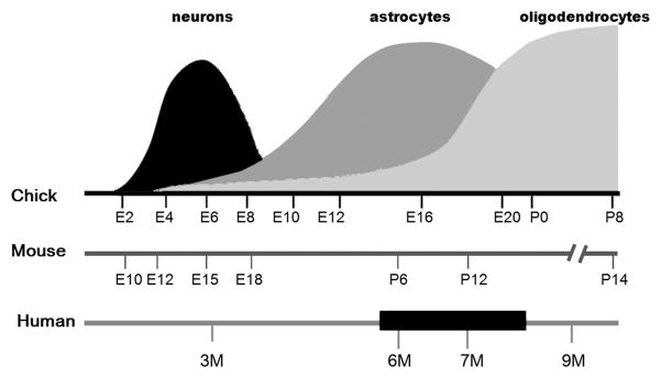
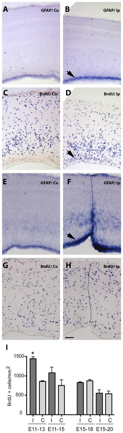
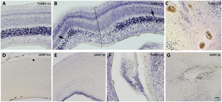
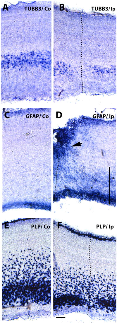
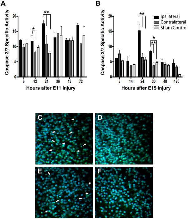
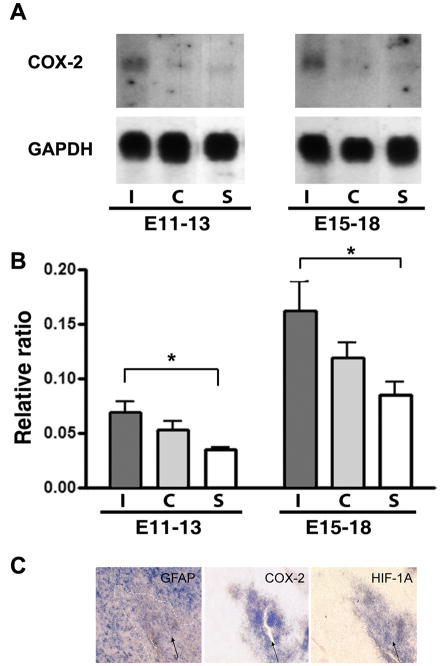
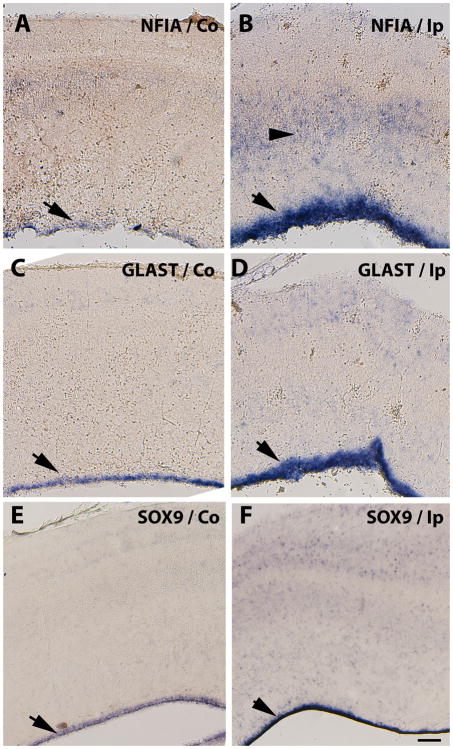
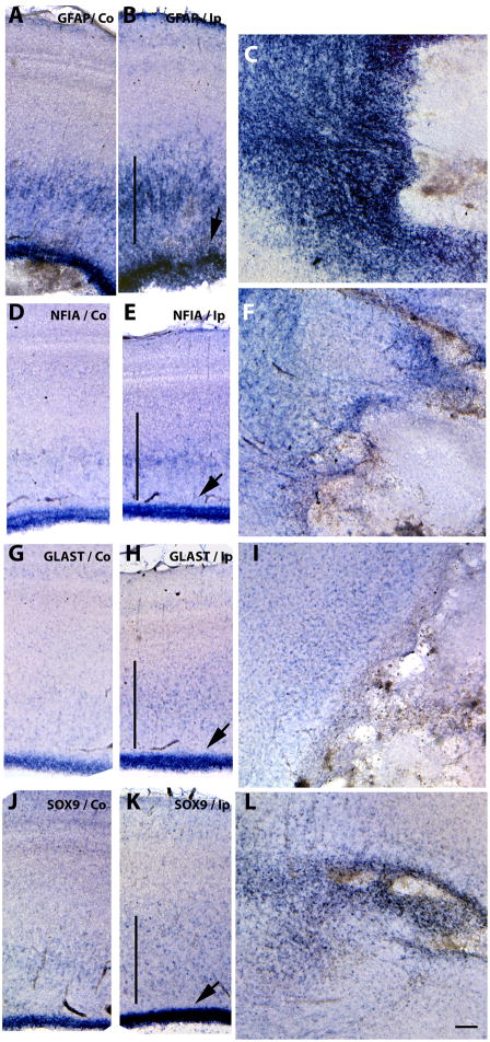
Penetrating traumatic insult during pregnancy is a leading cause of human fetal demise; in particular, trauma to the brain may lead to devastating long-term cognitive sequelae. Perinatal brain injury involves glial precursors, but the neural mechanisms controlling astrocyte ontogeny after injury remain incompletely understood, partly due to a lack of appropriate markers and animal models. We analyzed astrocyte precursor response to injury at the beginning (E11) and peak (E15) of gliogenesis in an avian tectal model of penetrating embryonic brain trauma, without confounding maternal and sibling effects. At both ages, lateral ventricular dilatation, necrotic foci, periventricular cysts and intraventricular hemorrhages were observed distal to stab wounds two days after a unilateral stab injury to optic tecta. Neuronal (TUBB3) and oligodendrocyte precursor (PLP) markers were down-regulated, even far-removed from the wound site. In contrast, the mature astrocyte marker, GFAP, was up-regulated at the wound site, around necrotic areas and cysts, plus in usual areas of GFAP expression. Increased inflammatory response and apoptotic cell death were also confirmed in the injured tecta. Increased expression of NFIA, SOX9 and GLAST at the wound site and in the ventricular zone (VZ) of the injured tecta indicated an astroglial precursor response. However, cell division increased in the VZ only in early (E11) injury, but not later (E15), indicating that in late injury the astrogliogenesis occurring after acute injury is predominantly due to precursor differentiation rather than precursor proliferation. The inability to replenish the glial precursor pool during the critical period of vulnerability to injury may be an important cause of subsequent developmental abnormalities.
怀孕期间穿透性创伤是导致人类胎儿死亡的主要原因;特别是大脑创伤可能导致毁灭性的长期认知后遗症。围产期脑损伤涉及神经胶质前体细胞,但控制损伤后星形胶质细胞发生的神经机制仍不完全清楚,部分原因是缺乏适当的标志物和动物模型。我们在穿透性胚胎脑创伤的禽类脑模型中分析了星形胶质细胞前体细胞对损伤的反应,在该模型中,在神经发生的早期(E11)和高峰期(E15),没有母体和兄弟姐妹的影响。在这两个年龄,在单侧视顶盖刺伤后两天,观察到侧脑室扩张、坏死灶、脑室周围囊肿和脑室内出血,这些损伤都位于刺伤部位的远端。神经元(TUBB3)和少突胶质前体细胞(PLP)标志物下调,甚至远离伤口部位。相比之下,成熟星形胶质细胞标志物 GFAP 在伤口部位、坏死区域和囊肿周围以及通常的 GFAP 表达区域上调。在受伤的视顶盖中还证实了炎症反应和凋亡细胞死亡增加。在受伤的视顶盖的伤口部位和脑室区(VZ),NFIA、SOX9 和 GLAST 的表达增加表明存在星形胶质前体细胞反应。然而,只有在早期(E11)损伤时,VZ 中的细胞分裂才增加,而在晚期(E15)损伤时则没有,这表明在晚期损伤中,急性损伤后发生的星形胶质发生主要是由于前体细胞分化而不是前体细胞增殖。在易受伤的关键时期,无法补充神经胶质前体细胞池,这可能是随后发育异常的一个重要原因。