Department of Animal Biology and Ecology, University of Cagliari, Via Ing. Tomaso Fiorelli, 09126 Cagliari, Italy,
Brain Struct Funct. 2011 Sep;216(3):171-82. doi: 10.1007/s00429-011-0312-2. Epub 2011 Apr 2.
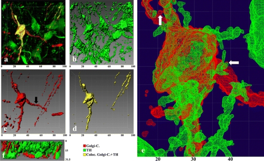
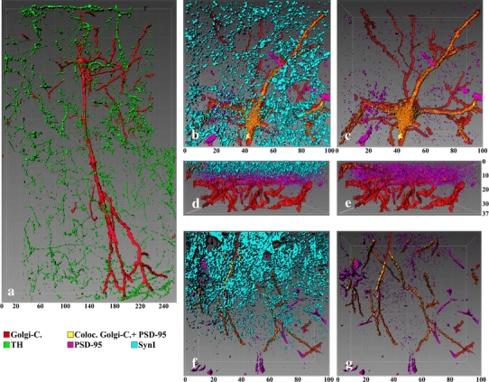
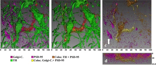
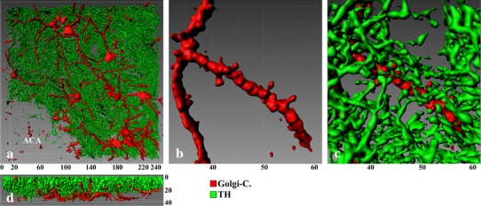
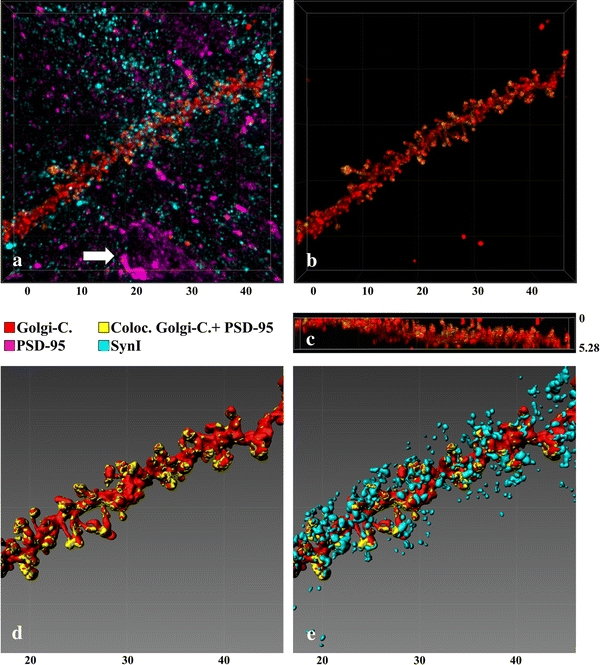
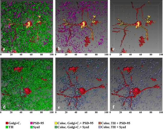
Visualization of neuronal elements is of fundamental importance in modern neuroscience. Golgi-Cox impregnation is a widely employed method that provides detailed information about morphological characteristics of neurons, but none regarding their neurochemical features. Immunocytochemical procedures, on the other hand, can provide a high degree of biochemical specificity but poorer morphological details, in particular if compared to Golgi-Cox impregnation. Hence, the combined use of these two approaches is highly desirable, especially for confocal microscopy that can exploit the advantages of both methods simultaneously. Here we show an innovative procedure of perfusion and fixation of brain tissue, that allows, by applying Golgi-Cox impregnation and immunofluorescence in the same histological section, to obtain high-quality histological material, with a very simple and inexpensive method. This procedure is based on three simple fixation steps: (1) a paraformaldehyde perfusion followed by a standard post-fixation to stabilize the subsequent immunofluorescence reaction; (2) the classical Golgi-Cox impregnation and (3) an immunofluorescence reaction in previously impregnated material. This combination allows simultaneous visualization of (a) the structural details (Golgi-Cox impregnated neurons), (b) the antigens' characterization, (c) the anatomical interactions between discrete neuronal elements and (d) the 3D reconstruction and modeling. The method is easy to perform and can be reproducibly applied by small laboratories and expanded through the use of different antibodies. Overall, the method presented in this study offers an innovative and powerful approach to study the nervous system, especially by using confocal microscopy.
神经元元素的可视化在现代神经科学中具有重要意义。高尔基-考克斯浸染是一种广泛应用的方法,它可以提供有关神经元形态特征的详细信息,但不能提供其神经化学特征的信息。免疫细胞化学程序可以提供高度的生化特异性,但形态细节较差,特别是与高尔基-考克斯浸染相比。因此,这两种方法的联合使用是非常理想的,特别是对于共聚焦显微镜,可以同时利用这两种方法的优点。在这里,我们展示了一种创新的脑组织灌注和固定程序,通过在同一组织切片上应用高尔基-考克斯浸染和免疫荧光,可以获得高质量的组织学材料,方法非常简单且经济实惠。该程序基于三个简单的固定步骤:(1)用多聚甲醛灌注,然后进行标准后固定,以稳定随后的免疫荧光反应;(2)经典的高尔基-考克斯浸染;(3)在先前浸染的材料中进行免疫荧光反应。这种组合允许同时观察(a)结构细节(高尔基-考克斯浸染的神经元),(b)抗原的特征,(c)离散神经元元素之间的解剖相互作用,以及(d)3D 重建和建模。该方法易于操作,可由小型实验室重复应用,并通过使用不同的抗体进行扩展。总的来说,本研究中提出的方法为研究神经系统提供了一种创新而强大的方法,特别是通过共聚焦显微镜。