• 文献检索
  • 文档翻译
  • 深度研究
  • 学术资讯
  • Suppr Zotero 插件Zotero 插件
  • 邀请有礼
  • 套餐&价格
  • 历史记录
应用&插件
Suppr Zotero 插件Zotero 插件浏览器插件Mac 客户端Windows 客户端微信小程序
定价
高级版会员购买积分包购买API积分包
服务
文献检索文档翻译深度研究API 文档MCP 服务
关于我们
关于 Suppr公司介绍联系我们用户协议隐私条款
关注我们

Suppr 超能文献

核心技术专利:CN118964589B侵权必究
粤ICP备2023148730 号-1Suppr @ 2026

文献检索

告别复杂PubMed语法,用中文像聊天一样搜索,搜遍4000万医学文献。AI智能推荐,让科研检索更轻松。

立即免费搜索

文件翻译

保留排版,准确专业,支持PDF/Word/PPT等文件格式,支持 12+语言互译。

免费翻译文档

深度研究

AI帮你快速写综述,25分钟生成高质量综述,智能提取关键信息,辅助科研写作。

立即免费体验

李斯特菌感染期间 IFN-β 的产生仅限于单核细胞/巨噬细胞谱系。

Production of IFN-β during Listeria monocytogenes infection is restricted to monocyte/macrophage lineage.

机构信息

Department of Molecular Biotechnology, Helmholtz Centre for Infection Research, Brunswick, Germany.

出版信息

PLoS One. 2011 Apr 11;6(4):e18543. doi: 10.1371/journal.pone.0018543.

DOI:10.1371/journal.pone.0018543
PMID:21494554
原文链接:https://pmc.ncbi.nlm.nih.gov/articles/PMC3073975/
Abstract

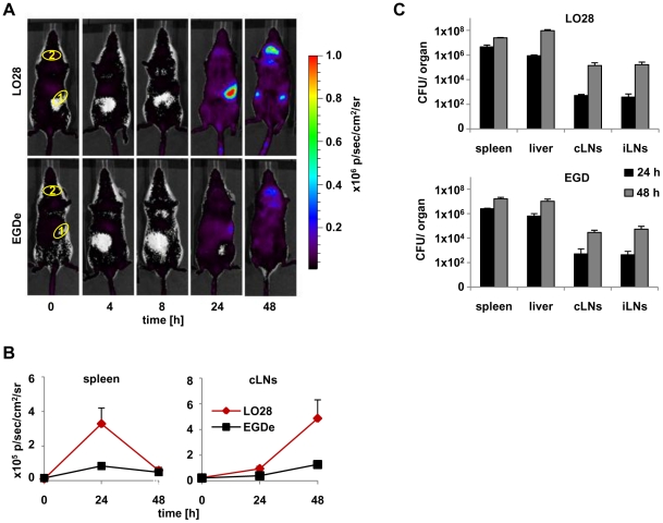
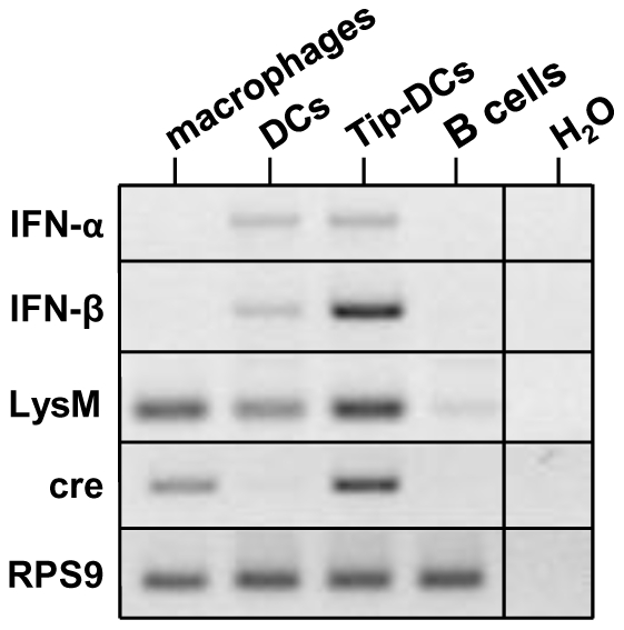
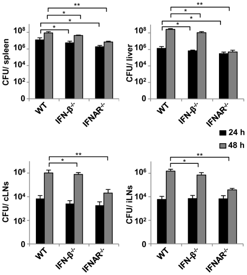
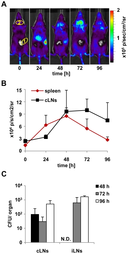
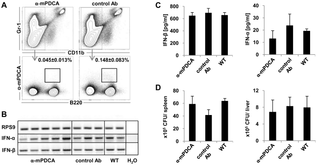
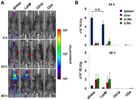
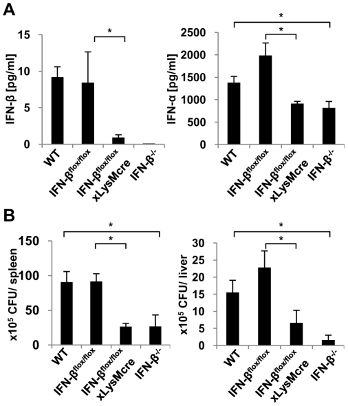
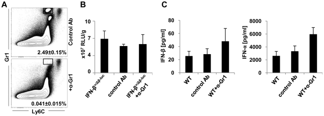
The family of type I interferons (IFN), which consists of several IFN-α and one IFN-β, are produced not only after stimulation by viruses, but also after infection with non-viral pathogens. In the course of bacterial infections, these cytokines could be beneficial or detrimental. IFN-β is the primary member of type I IFN that initiates a cascade of IFN-α production. Here we addressed the question which cells are responsible for IFN-β expression after infection with the intracellular pathogen Listeria monocytogenes by using a genetic approach. By means of newly established reporter mice, maximum of IFN-β expression was observed at 24 hours post infection in spleen and, surprisingly, 48 hours post infection in colonized cervical and inguinal lymph nodes. Colonization of lymph nodes was independent of the type I IFN signaling, as well as bacterial dose and strain. Using cell specific reporter function and conditional deletions we could define cells expressing LysM as the major IFN-β producers, with cells formerly defined as Tip-DCs being the highest. Neutrophilic granulocytes, dendritic cells and plasmacytoid dendritic cells did not significantly contribute to type I IFN production.

摘要

I 型干扰素(IFN)家族由几种 IFN-α 和一种 IFN-β 组成,不仅在病毒刺激后产生,而且在非病毒病原体感染后也会产生。在细菌感染过程中,这些细胞因子可能有益也可能有害。IFN-β 是启动 IFN-α 产生级联反应的 I 型 IFN 的主要成员。在这里,我们通过遗传方法解决了感染细胞内病原体李斯特菌后哪些细胞负责 IFN-β 表达的问题。通过新建立的报告小鼠,在感染后 24 小时观察到脾脏中 IFN-β 的最大表达,令人惊讶的是,在感染后 48 小时在定植的颈淋巴结和腹股沟淋巴结中观察到 IFN-β 的最大表达。淋巴结的定植不依赖于 I 型 IFN 信号,也不依赖于细菌剂量和菌株。使用细胞特异性报告功能和条件性缺失,我们可以定义表达 LysM 的细胞为主要的 IFN-β 产生细胞,其中以前定义为 Tip-DC 的细胞表达最高。中性粒细胞、树突状细胞和浆细胞样树突状细胞对 I 型 IFN 产生没有显著贡献。

https://cdn.ncbi.nlm.nih.gov/pmc/blobs/b8f0/3073975/08ceab11fb46/pone.0018543.g008.jpg
https://cdn.ncbi.nlm.nih.gov/pmc/blobs/b8f0/3073975/4652ca6cde99/pone.0018543.g001.jpg
https://cdn.ncbi.nlm.nih.gov/pmc/blobs/b8f0/3073975/35e680d1771c/pone.0018543.g002.jpg
https://cdn.ncbi.nlm.nih.gov/pmc/blobs/b8f0/3073975/fdba295cd3e5/pone.0018543.g003.jpg
https://cdn.ncbi.nlm.nih.gov/pmc/blobs/b8f0/3073975/af0c82fb641f/pone.0018543.g004.jpg
https://cdn.ncbi.nlm.nih.gov/pmc/blobs/b8f0/3073975/3d23a0744586/pone.0018543.g005.jpg
https://cdn.ncbi.nlm.nih.gov/pmc/blobs/b8f0/3073975/3e2c4325ef89/pone.0018543.g006.jpg
https://cdn.ncbi.nlm.nih.gov/pmc/blobs/b8f0/3073975/5d8c1cbd8826/pone.0018543.g007.jpg
https://cdn.ncbi.nlm.nih.gov/pmc/blobs/b8f0/3073975/08ceab11fb46/pone.0018543.g008.jpg

相似文献

1
Production of IFN-β during Listeria monocytogenes infection is restricted to monocyte/macrophage lineage.李斯特菌感染期间 IFN-β 的产生仅限于单核细胞/巨噬细胞谱系。
PLoS One. 2011 Apr 11;6(4):e18543. doi: 10.1371/journal.pone.0018543.
2
Type I IFN Does Not Promote Susceptibility to Foodborne Listeria monocytogenes.I型干扰素不会增加对食源性单核细胞增生李斯特菌的易感性。
J Immunol. 2016 Apr 1;196(7):3109-16. doi: 10.4049/jimmunol.1502192. Epub 2016 Feb 19.
3
Characterization of the interferon-producing cell in mice infected with Listeria monocytogenes.感染单核细胞增生李斯特菌的小鼠中产生干扰素细胞的特征分析。
PLoS Pathog. 2009 Mar;5(3):e1000355. doi: 10.1371/journal.ppat.1000355. Epub 2009 Mar 27.
4
A fluorescence reporter model defines "Tip-DCs" as the cellular source of interferon β in murine listeriosis.荧光报告模型将“Tip-DCs”定义为鼠李斯特菌病中干扰素 β 的细胞来源。
PLoS One. 2010 Dec 16;5(12):e15567. doi: 10.1371/journal.pone.0015567.
5
Plasmacytoid dendritic cells mature independently of MyD88 and IFN-αβR in response to Listeria and stimulate CD8 T cells.浆细胞样树突状细胞在应对李斯特菌时独立于 MyD88 和 IFN-αβR 成熟,并刺激 CD8 T 细胞。
Immunol Lett. 2011 Aug 30;138(2):104-12. doi: 10.1016/j.imlet.2011.03.007. Epub 2011 Mar 29.
6
The timing of IFNβ production affects early innate responses to Listeria monocytogenes and determines the overall outcome of lethal infection.IFNβ 的产生时间会影响李斯特菌感染早期的固有免疫反应,并决定致死性感染的整体结局。
PLoS One. 2012;7(8):e43455. doi: 10.1371/journal.pone.0043455. Epub 2012 Aug 17.
7
IFN-β, but not IFN-α, is Responsible for the Pro-Bacterial Effect of Type I Interferon.IFN-β,而不是 IFN-α,是造成 I 型干扰素的促细菌作用的原因。
Cell Physiol Biochem. 2021 May 14;55(3):256-264. doi: 10.33594/000000370.
8
IFN-γ protects from apoptotic neutrophil-mediated tissue injury during acute Listeria monocytogenes infection.IFN-γ 可防止急性李斯特菌感染期间中性粒细胞介导的细胞凋亡性组织损伤。
Eur J Immunol. 2018 Sep;48(9):1470-1480. doi: 10.1002/eji.201847491. Epub 2018 Jul 25.
9
Sources of interferon-gamma (IFN-gamma) in early immune response to Listeria monocytogenes.单核细胞增生李斯特菌早期免疫反应中γ-干扰素(IFN-γ)的来源
Immunobiology. 2005;210(9):673-83. doi: 10.1016/j.imbio.2005.07.003.
10
Listeria monocytogenes-induced gamma interferon secretion by intestinal intraepithelial gamma/delta T lymphocytes.单核细胞增生李斯特菌诱导肠道上皮γ/δ T淋巴细胞分泌γ干扰素
Infect Immun. 1993 May;61(5):2154-61. doi: 10.1128/iai.61.5.2154-2161.1993.

引用本文的文献

1
Type I Interferon, Induced by Adenovirus or Adenoviral Vector Infection, Regulates the Cytokine Response to Lipopolysaccharide in a Macrophage Type-Specific Manner.由腺病毒或腺病毒载体感染诱导产生的I型干扰素,以巨噬细胞类型特异性方式调节对脂多糖的细胞因子反应。
J Innate Immun. 2024;16(1):226-247. doi: 10.1159/000538282. Epub 2024 Mar 25.
2
Brain endothelial STING1 activation by -sequestered heme promotes cerebral malaria via type I IFN response.被 - 隔离的血红素激活脑内皮细胞 STING1 促进脑型疟疾通过 I 型干扰素反应。
Proc Natl Acad Sci U S A. 2022 Sep 6;119(36):e2206327119. doi: 10.1073/pnas.2206327119. Epub 2022 Aug 29.
3

本文引用的文献

1
A fluorescence reporter model defines "Tip-DCs" as the cellular source of interferon β in murine listeriosis.荧光报告模型将“Tip-DCs”定义为鼠李斯特菌病中干扰素 β 的细胞来源。
PLoS One. 2010 Dec 16;5(12):e15567. doi: 10.1371/journal.pone.0015567.
2
Antagonistic crosstalk between type I and II interferons and increased host susceptibility to bacterial infections.I 型和 II 型干扰素之间的拮抗串扰和宿主对细菌感染易感性的增加。
Virulence. 2010 Sep-Oct;1(5):418-22. doi: 10.4161/viru.1.5.12787.
3
Applications of myeloid-specific promoters in transgenic mice support in vivo imaging and functional genomics but do not support the concept of distinct macrophage and dendritic cell lineages or roles in immunity.
The use of foodborne infection to evaluate bacterial pathogenesis and host response.
利用食源性病原体感染来评估细菌发病机制和宿主反应。
Methods Cell Biol. 2022;168:299-314. doi: 10.1016/bs.mcb.2021.12.020. Epub 2022 Jan 29.
4
Myeloid Cell CK2 Regulates Inflammation and Resistance to Bacterial Infection.髓系细胞 CK2 调节炎症反应和抵抗细菌感染。
Front Immunol. 2020 Dec 8;11:590266. doi: 10.3389/fimmu.2020.590266. eCollection 2020.
5
PD-L1 Reverse Signaling in Dermal Dendritic Cells Promotes Dendritic Cell Migration Required for Skin Immunity.PD-L1 信号在皮肤树突状细胞中的反向传递促进了树突状细胞的迁移,这是皮肤免疫所必需的。
Cell Rep. 2020 Oct 13;33(2):108258. doi: 10.1016/j.celrep.2020.108258.
6
Plasmacytoid dendritic cells in the eye.眼内浆细胞样树突状细胞。
Prog Retin Eye Res. 2021 Jan;80:100877. doi: 10.1016/j.preteyeres.2020.100877. Epub 2020 Jul 24.
7
Production of IFNβ by Conventional Dendritic Cells after Stimulation with Viral Compounds and IFNβ-Independent IFNAR1-Signaling Pathways are Associated with Aggravation of Polymicrobial Sepsis.病毒化合物刺激后常规树突状细胞产生 IFNβ 和 IFNβ 非依赖的 IFNAR1 信号通路与多微生物脓毒症的加重有关。
Int J Mol Sci. 2019 Sep 7;20(18):4410. doi: 10.3390/ijms20184410.
8
Innate and Adaptive Immune Responses during Infection.感染期间的先天和适应性免疫反应。
Microbiol Spectr. 2019 May;7(3). doi: 10.1128/microbiolspec.GPP3-0065-2019.
9
Sources of Type I Interferons in Infectious Immunity: Plasmacytoid Dendritic Cells Not Always in the Driver's Seat.固有免疫中Ⅰ型干扰素的来源:浆细胞样树突状细胞并非一直占据主导地位。
Front Immunol. 2019 Apr 12;10:778. doi: 10.3389/fimmu.2019.00778. eCollection 2019.
10
ISRE-Reporter Mouse Reveals High Basal and Induced Type I IFN Responses in Inflammatory Monocytes.ISRE-Reporter 小鼠揭示了炎性单核细胞中高基础和诱导的 I 型 IFN 反应。
Cell Rep. 2018 Dec 4;25(10):2784-2796.e3. doi: 10.1016/j.celrep.2018.11.030.
髓系特异性启动子在转基因小鼠中的应用支持体内成像和功能基因组学,但不支持巨噬细胞和树突状细胞谱系或在免疫中作用的独特概念。
J Leukoc Biol. 2011 Apr;89(4):525-38. doi: 10.1189/jlb.0810472. Epub 2010 Dec 17.
4
Hepatitis B virus: from diagnosis to treatment.乙型肝炎病毒:从诊断到治疗
Pathol Biol (Paris). 2010 Aug;58(4):245-53. doi: 10.1016/j.patbio.2010.05.002. Epub 2010 Jul 1.
5
c-di-AMP secreted by intracellular Listeria monocytogenes activates a host type I interferon response.细胞内李斯特菌分泌的 c-di-AMP 激活宿主Ⅰ型干扰素应答。
Science. 2010 Jun 25;328(5986):1703-5. doi: 10.1126/science.1189801. Epub 2010 May 27.
6
Neutrophils responsive to endogenous IFN-beta regulate tumor angiogenesis and growth in a mouse tumor model.内源性 IFN-β 反应性中性粒细胞调节小鼠肿瘤模型中的肿瘤血管生成和生长。
J Clin Invest. 2010 Apr;120(4):1151-64. doi: 10.1172/JCI37223. Epub 2010 Mar 8.
7
Development of monocytes, macrophages, and dendritic cells.单核细胞、巨噬细胞和树突状细胞的发育。
Science. 2010 Feb 5;327(5966):656-61. doi: 10.1126/science.1178331.
8
Induction of IFN-alphabeta enables Listeria monocytogenes to suppress macrophage activation by IFN-gamma.IFN-αβ 的诱导使李斯特菌能够抑制 IFN-γ 诱导的巨噬细胞活化。
J Exp Med. 2010 Feb 15;207(2):327-37. doi: 10.1084/jem.20091746. Epub 2010 Feb 1.
9
Interferon-beta therapy for multiple sclerosis--is the injection site the relevant action site?β-干扰素治疗多发性硬化症——注射部位是相关作用部位吗?
J Neuroimmunol. 2009 Oct 30;215(1-2):117-21. doi: 10.1016/j.jneuroim.2009.08.011. Epub 2009 Sep 20.
10
Intracellular innate immune cascades and interferon defenses that control hepatitis C virus.细胞内先天免疫级联反应和干扰素防御系统控制丙型肝炎病毒。
J Interferon Cytokine Res. 2009 Sep;29(9):489-98. doi: 10.1089/jir.2009.0063.