Institute of Clinical Pharmacology, Hannover Medical School, Hannover, Germany.
PLoS One. 2011 Apr 8;6(4):e18737. doi: 10.1371/journal.pone.0018737.
Ozone concentrations in ambient air are related to cardiopulmonary perturbations in the aging population. Increased central sympathetic nerve activity induced by local airway inflammation may be one possible mechanism.
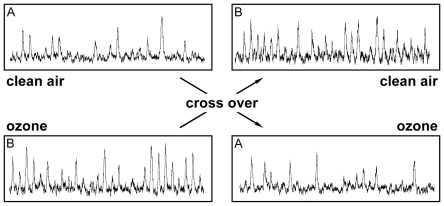
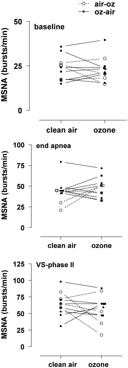
METHODOLOGY/PRINCIPAL FINDINGS: To elucidate this issue further, we performed a randomized, double-blind, cross-over study, including 14 healthy subjects (3 females, age 22-47 years), who underwent a 3 h exposure with intermittent exercise to either ozone (250 ppb) or clean air. Induced sputum was collected 3 h after exposure. Nineteen to 22 hours after exposure, we recorded ECG, finger blood pressure, brachial blood pressure, respiration, cardiac output, and muscle sympathetic nerve activity (MSNA) at rest, during deep breathing, maximum-inspiratory breath hold, and a Valsalva maneuver. While the ozone exposure induced the expected airway inflammation, as indicated by a significant increase in sputum neutrophils, we did not detect a significant estimated treatment effect adjusted for period on cardiovascular measurements. Resting heart rate (clean air: 59±2, ozone 60±2 bpm), blood pressure (clean air: 121±3/71±2 mmHg; ozone: 121±2/71±2 mmHg), cardiac output (clean air: 7.42±0.29 mmHg; ozone: 7.98±0.60 l/min), and plasma norepinephrine levels (clean air: 213±21 pg/ml; ozone: 202±16 pg/ml), were similar on both study days. No difference of resting MSNA was observed between ozone and air exposure (air: 23±2, ozone: 23±2 bursts/min). Maximum MSNA obtained at the end of apnea (air: 44±4, ozone: 48±4 bursts/min) and during the phase II of the Valsalva maneuver (air: 64±5, ozone: 57±6 bursts/min) was similar.
CONCLUSIONS/SIGNIFICANCE: Our study suggests that acute ozone-induced airway inflammation does not increase resting sympathetic nerve traffic in healthy subjects, an observation that is relevant for environmental health. However, we can not exclude that chronic airway inflammation may contribute to sympathetic activation.
环境空气中的臭氧浓度与老年人群中心肺功能紊乱有关。局部气道炎症引起的中枢交感神经活性增加可能是一种可能的机制。
方法/主要发现:为了进一步阐明这个问题,我们进行了一项随机、双盲、交叉研究,包括 14 名健康受试者(3 名女性,年龄 22-47 岁),他们在 3 小时内间歇性运动暴露于臭氧(250ppb)或清洁空气。暴露后 3 小时采集诱导痰。暴露后 19-22 小时,我们在休息、深呼吸、最大吸气屏气和瓦尔萨尔瓦动作时记录心电图、手指血压、肱动脉血压、呼吸、心输出量和肌肉交感神经活动(MSNA)。虽然臭氧暴露引起了预期的气道炎症,表现为痰中性粒细胞显著增加,但我们没有发现心血管测量的预期治疗效果有显著差异。静息心率(清洁空气:59±2,臭氧 60±2bpm)、血压(清洁空气:121±3/71±2mmHg;臭氧:121±2/71±2mmHg)、心输出量(清洁空气:7.42±0.29mmHg;臭氧:7.98±0.60l/min)和血浆去甲肾上腺素水平(清洁空气:213±21pg/ml;臭氧:202±16pg/ml)在两天的研究中相似。在臭氧和空气暴露之间,静息 MSNA 没有差异(空气:23±2,臭氧:23±2 脉冲/分钟)。在最大吸气屏气结束时(空气:44±4,臭氧:48±4 脉冲/分钟)和瓦尔萨尔瓦动作第二期(空气:64±5,臭氧:57±6 脉冲/分钟)时获得的最大 MSNA 相似。
结论/意义:我们的研究表明,急性臭氧诱导的气道炎症不会增加健康受试者的静息交感神经活动,这一观察结果与环境卫生有关。然而,我们不能排除慢性气道炎症可能导致交感神经激活。