Department of Neurophysiology and Pharmacology, Center of Physiology and Pharmacology, Medical University of Vienna, Vienna, Austria.
Br J Pharmacol. 2011 Nov;164(5):1522-33. doi: 10.1111/j.1476-5381.2011.01466.x.
P2Y(1) , P2Y(2) , P2Y(4) , P2Y(12) and P2Y(13) receptors for nucleotides have been reported to mediate presynaptic inhibition, but unequivocal evidence for facilitatory presynaptic P2Y receptors is not available. The search for such receptors was the purpose of this study.
In primary cultures of rat superior cervical ganglion neurons and in PC12 cell cultures, currents were recorded via the perforated patch clamp technique, and the release of [(3) H]-noradrenaline was determined.
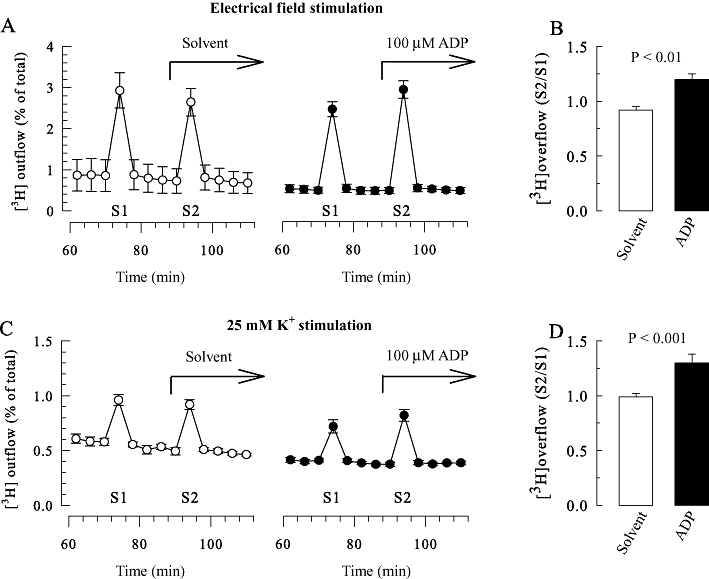
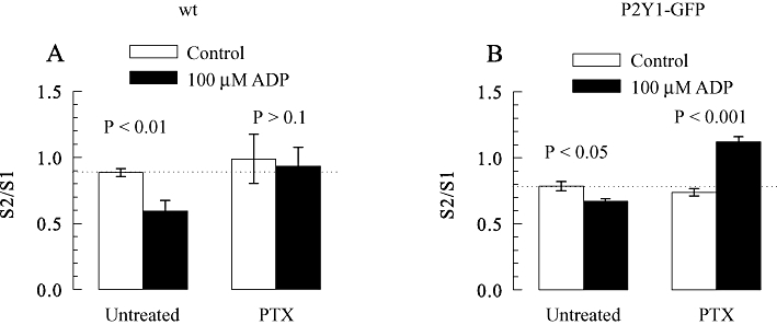
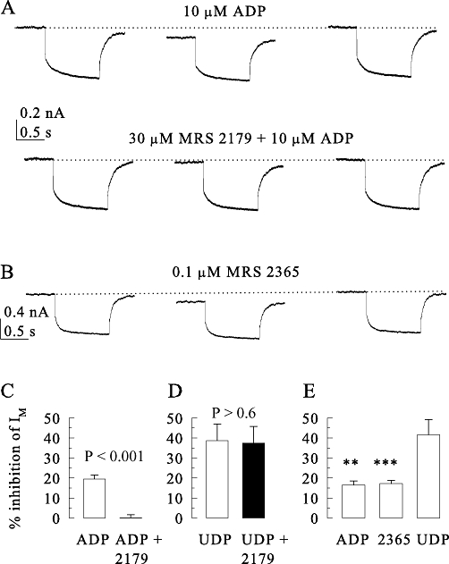
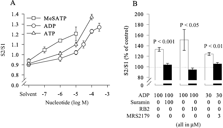
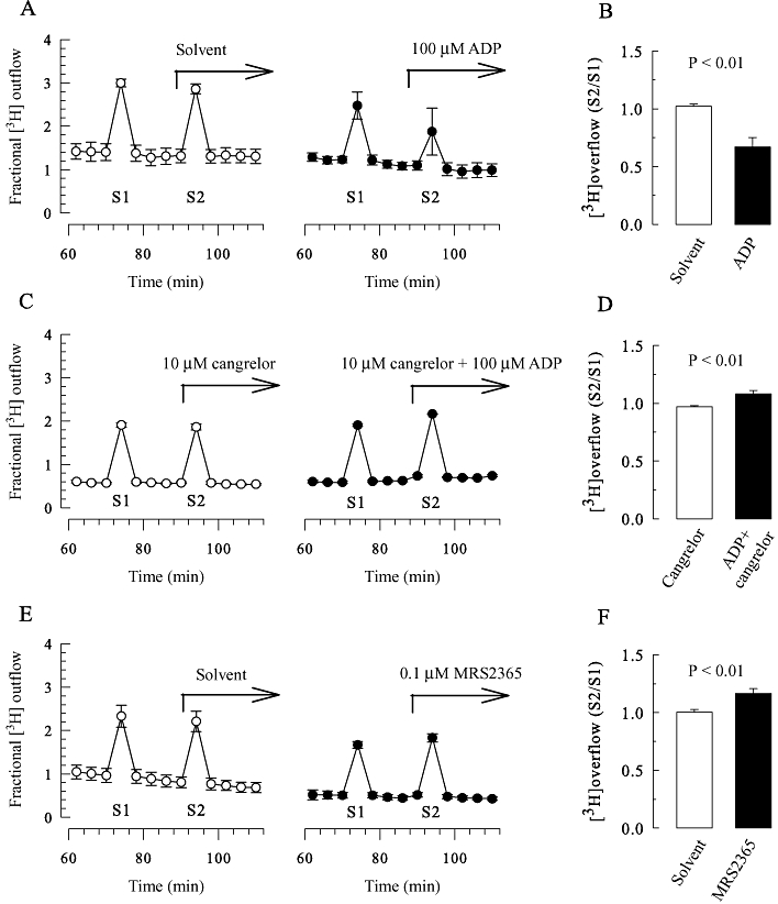
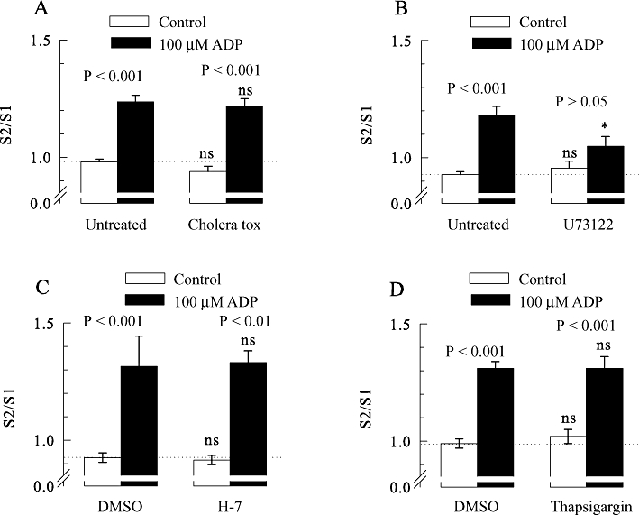
ADP, 2-methylthio-ATP and ATP enhanced stimulation-evoked (3) H overflow from superior cervical ganglion neurons, treated with pertussis toxin to prevent the signalling of inhibitory G proteins. This effect was abolished by P2Y(1) antagonists and by inhibition of phospholipase C, but not by inhibition of protein kinase C or depletion of intracellular Ca(2+) stores. ADP and a specific P2Y(1) agonist caused inhibition of Kv7 channels, and this was prevented by a respective antagonist. In neurons not treated with pertussis toxin, (3) H overflow was also enhanced by a specific P2Y(1) agonist and by ADP, but only when the P2Y(12) receptors were blocked. ADP also enhanced K(+) -evoked (3) H overflow from PC12 cells treated with pertussis toxin, but only in a clone expressing recombinant P2Y(1) receptors.
These results demonstrate that presynaptic P2Y(1) receptors mediate facilitation of transmitter release from sympathetic neurons most likely through inhibition of Kv7 channels.
已报道核苷酸的 P2Y(1)、P2Y(2)、P2Y(4)、P2Y(12)和 P2Y(13)受体可介导突触前抑制,但尚无促进突触前 P2Y 受体的明确证据。本研究旨在寻找此类受体。
在大鼠颈上交感神经节神经元的原代培养物和 PC12 细胞培养物中,通过穿孔膜片钳技术记录电流,并测定 [(3)H]-去甲肾上腺素的释放。
ADP、2-甲基硫代-ATP 和 ATP 增强了用百日咳毒素处理以防止抑制性 G 蛋白信号传递的颈上交感神经节神经元刺激诱发的 [(3)H]溢出。这种效应被 P2Y(1)拮抗剂和磷脂酶 C 抑制所消除,但不受蛋白激酶 C 抑制或细胞内 Ca(2+)储存耗竭的影响。ADP 和一种特异性 P2Y(1)激动剂引起 Kv7 通道抑制,而这一效应被相应的拮抗剂所阻止。在未经百日咳毒素处理的神经元中,(3)H 溢出也被一种特异性 P2Y(1)激动剂和 ADP 增强,但只有当 P2Y(12)受体被阻断时才会增强。ADP 还增强了用百日咳毒素处理的表达重组 P2Y(1)受体的 PC12 细胞中 K(+)诱发的 [(3)H]溢出。
这些结果表明,突触前 P2Y(1)受体通过抑制 Kv7 通道介导交感神经元递质释放的易化,最有可能通过抑制 Kv7 通道介导。