• 文献检索
  • 文档翻译
  • 深度研究
  • 学术资讯
  • Suppr Zotero 插件Zotero 插件
  • 邀请有礼
  • 套餐&价格
  • 历史记录
应用&插件
Suppr Zotero 插件Zotero 插件浏览器插件Mac 客户端Windows 客户端微信小程序
定价
高级版会员购买积分包购买API积分包
服务
文献检索文档翻译深度研究API 文档MCP 服务
关于我们
关于 Suppr公司介绍联系我们用户协议隐私条款
关注我们

Suppr 超能文献

核心技术专利:CN118964589B侵权必究
粤ICP备2023148730 号-1Suppr @ 2026

文献检索

告别复杂PubMed语法,用中文像聊天一样搜索,搜遍4000万医学文献。AI智能推荐,让科研检索更轻松。

立即免费搜索

文件翻译

保留排版,准确专业,支持PDF/Word/PPT等文件格式,支持 12+语言互译。

免费翻译文档

深度研究

AI帮你快速写综述,25分钟生成高质量综述,智能提取关键信息,辅助科研写作。

立即免费体验

CT 结肠成像时同时进行骨质疏松症筛查:使用 MDCT 衰减技术评估骨密度与 DXA 参考标准比较。

Simultaneous screening for osteoporosis at CT colonography: bone mineral density assessment using MDCT attenuation techniques compared with the DXA reference standard.

机构信息

University of Wisconsin School of Medicine and Public Health, Madison, WI 53792-3252, USA.

出版信息

J Bone Miner Res. 2011 Sep;26(9):2194-203. doi: 10.1002/jbmr.428.

DOI:10.1002/jbmr.428
PMID:21590738
原文链接:https://pmc.ncbi.nlm.nih.gov/articles/PMC3304444/
Abstract

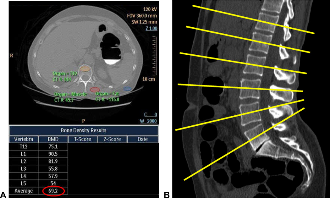
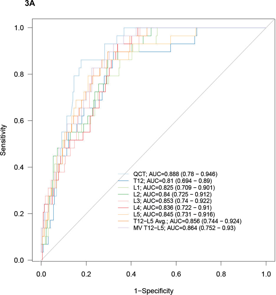
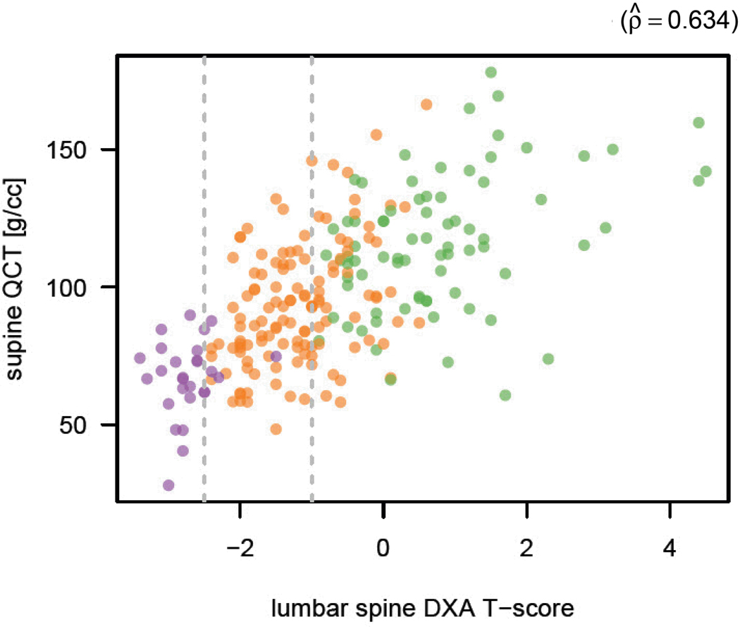
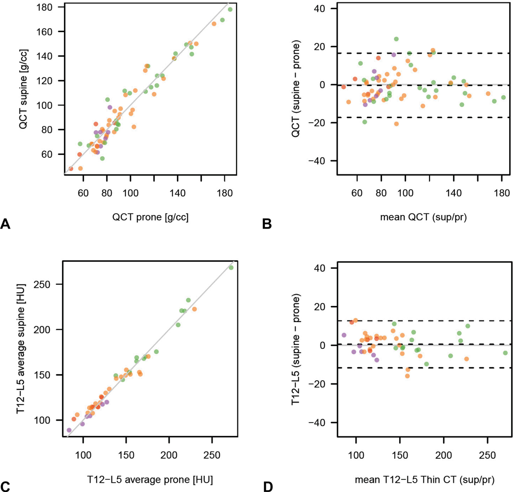
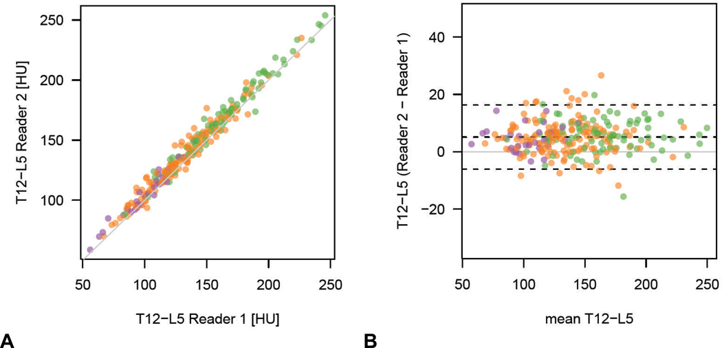
The purpose of this study was to evaluate the utility of lumbar spine attenuation measurement for bone mineral density (BMD) assessment at screening computed tomographic colonography (CTC) using central dual-energy X-ray absorptiometry (DXA) as the reference standard. Two-hundred and fifty-two adults (240 women and 12 men; mean age 58.9 years) underwent CTC screening and central DXA BMD measurement within 2 months (mean interval 25.0 days). The lowest DXA T-score between the spine and hip served as the reference standard, with low BMD defined per World Health Organization as osteoporosis (DXA T-score ≤ -2.5) or osteopenia (DXA T-score between -1.0 and -2.4). Both phantomless quantitative computed tomography (QCT) and simple nonangled region-of-interest (ROI) multi-detector CT (MDCT) attenuation measurements were applied to the T(12) -L(5) levels. The ability to predict osteoporosis and low BMD (osteoporosis or osteopenia) by DXA was assessed. A BMD cut-off of 90 mg/mL at phantomless QCT yielded 100% sensitivity for osteoporosis (29 of 29) and a specificity of 63.8% (143 of 224); 87.2% (96 of 110) below this threshold had low BMD and 49.6% (69 of 139) above this threshold had normal BMD at DXA. At L(1) , a trabecular ROI attenuation cut-off of 160 HU was 100% sensitive for osteoporosis (29 of 29), with a specificity of 46.4% (104 of 224); 83.9% (125 of 149) below this threshold had low BMD and 57.5% (59/103) above had normal BMD at DXA. ROI performance was similar at all individual T(12) -L(5) levels. At ROC analysis, AUC for osteoporosis was 0.888 for phantomless QCT [95% confidence interval (CI) 0.780-0.946] and ranged from 0.825 to 0.853 using trabecular ROIs at single lumbar levels (0.864; 95% CI 0.752-0.930 at multivariate analysis). Supine-prone reproducibility was better with the simple ROI method compared with QCT. It is concluded that both phantomless QCT and simple ROI attenuation measurements of the lumbar spine are effective for BMD screening at CTC with high sensitivity for osteoporosis, as defined by the DXA T-score.

摘要

本研究旨在评估腰椎衰减测量在使用中央双能 X 射线吸收法(DXA)作为参考标准的筛查性计算机断层结肠造影术(CTC)中的骨密度(BMD)评估中的效用。252 名成年人(240 名女性和 12 名男性;平均年龄 58.9 岁)在 2 个月内接受 CTC 筛查和中央 DXA BMD 测量(平均间隔 25.0 天)。最低的 DXA T 评分(脊柱和臀部之间)用作参考标准,根据世界卫生组织的定义,低 BMD 为骨质疏松症(DXA T 评分≤-2.5)或骨量减少症(DXA T 评分在-1.0 至-2.4 之间)。均采用无伪影定量 CT(QCT)和简单非角度感兴趣区(ROI)多探测器 CT(MDCT)衰减测量法对 T(12)-L(5)水平进行评估。评估了 DXA 预测骨质疏松症和低 BMD(骨质疏松症或骨量减少症)的能力。无伪影 QCT 中 BMD 截断值为 90mg/mL 时,对骨质疏松症的敏感性为 100%(29/29),特异性为 63.8%(143/224);低于此阈值的 87.2%(110/125)有低 BMD,高于此阈值的 49.6%(139/279)在 DXA 中有正常的 BMD。在 L(1),骨小梁 ROI 衰减截断值为 160HU 时,对骨质疏松症的敏感性为 100%(29/29),特异性为 46.4%(104/224);低于此阈值的 83.9%(149/178)有低 BMD,高于此阈值的 57.5%(103/181)在 DXA 中有正常的 BMD。ROI 性能在所有单独的 T(12)-L(5)水平上均相似。在 ROC 分析中,无伪影 QCT 的骨质疏松症 AUC 为 0.888(95%置信区间[CI]为 0.780-0.946),在单个腰椎水平使用骨小梁 ROI 时范围为 0.825 至 0.853(0.864;95%CI 为 0.752-0.930 在多变量分析中)。与 QCT 相比,仰卧位-俯卧位重复性更好。结论是,无伪影 QCT 和腰椎简单 ROI 衰减测量均能有效筛查 CTC 的 BMD,对骨质疏松症具有较高的敏感性,其定义为 DXA T 评分。

https://cdn.ncbi.nlm.nih.gov/pmc/blobs/aaff/3304444/09ac35671418/nihms302217f7.jpg
https://cdn.ncbi.nlm.nih.gov/pmc/blobs/aaff/3304444/cd0d86043ad9/nihms302217f1a.jpg
https://cdn.ncbi.nlm.nih.gov/pmc/blobs/aaff/3304444/07847c92d476/nihms302217f2.jpg
https://cdn.ncbi.nlm.nih.gov/pmc/blobs/aaff/3304444/59e031384240/nihms302217f3a.jpg
https://cdn.ncbi.nlm.nih.gov/pmc/blobs/aaff/3304444/43ce16f515f5/nihms302217f4.jpg
https://cdn.ncbi.nlm.nih.gov/pmc/blobs/aaff/3304444/9e8caf4332e1/nihms302217f5.jpg
https://cdn.ncbi.nlm.nih.gov/pmc/blobs/aaff/3304444/497e5c4f0dd0/nihms302217f6.jpg
https://cdn.ncbi.nlm.nih.gov/pmc/blobs/aaff/3304444/09ac35671418/nihms302217f7.jpg

相似文献

1
Simultaneous screening for osteoporosis at CT colonography: bone mineral density assessment using MDCT attenuation techniques compared with the DXA reference standard.CT 结肠成像时同时进行骨质疏松症筛查:使用 MDCT 衰减技术评估骨密度与 DXA 参考标准比较。
J Bone Miner Res. 2011 Sep;26(9):2194-203. doi: 10.1002/jbmr.428.
2
Vertebral bone attenuation on low-dose chest CT: quantitative volumetric analysis for bone fragility assessment.低剂量胸部CT上的椎体骨质衰减:用于评估骨脆性的定量容积分析
Osteoporos Int. 2017 Jan;28(1):329-338. doi: 10.1007/s00198-016-3724-2. Epub 2016 Aug 1.
3
Discordance in lumbar bone mineral density measurements by quantitative computed tomography and dual-energy X-ray absorptiometry in postmenopausal women: a prospective comparative study.绝经后妇女定量计算机断层扫描和双能 X 射线吸收法测定腰椎骨密度的不相符:一项前瞻性对比研究。
Spine J. 2023 Feb;23(2):295-304. doi: 10.1016/j.spinee.2022.10.014. Epub 2022 Nov 4.
4
Diagnostic accuracy of quantitative dual-energy CT-based bone mineral density assessment in comparison to Hounsfield unit measurements using dual x-ray absorptiometry as standard of reference.定量双能 CT 骨密度评估与双能 X 射线吸收法作为参考标准的 Hounsfield 单位测量的诊断准确性比较。
Eur J Radiol. 2020 Nov;132:109321. doi: 10.1016/j.ejrad.2020.109321. Epub 2020 Sep 28.
5
Quantitative computed tomography has higher sensitivity detecting critical bone mineral density compared to dual-energy X-ray absorptiometry in postmenopausal women and elderly men with osteoporotic fractures: a real-life study.定量计算机断层扫描在检测绝经后妇女和老年骨质疏松性骨折男性的临界骨密度方面比双能 X 射线吸收法更敏感:一项真实研究。
Arch Orthop Trauma Surg. 2024 Jan;144(1):179-188. doi: 10.1007/s00402-023-05070-y. Epub 2023 Oct 5.
6
Comparison of femoral neck BMD evaluation obtained using Lunar DXA and QCT with asynchronous calibration from CT colonography.比较使用 Lunar DXA 和 QCT 与 CT 结肠成像的异步校准获得的股骨颈 BMD 评估。
J Clin Densitom. 2015 Jan-Mar;18(1):5-12. doi: 10.1016/j.jocd.2014.03.002. Epub 2014 May 28.
7
Opportunistic screening for osteoporosis by routine CT in Southern Europe.在南欧,通过常规 CT 进行骨质疏松症的机会性筛查。
Osteoporos Int. 2017 Mar;28(3):983-990. doi: 10.1007/s00198-016-3804-3. Epub 2017 Jan 20.
8
Effect of IV contrast on lumbar trabecular attenuation at routine abdominal CT: correlation with DXA and implications for opportunistic osteoporosis screening.静脉内造影剂对常规腹部CT检查时腰椎小梁骨密度的影响:与双能X线吸收法的相关性及对机会性骨质疏松症筛查的意义
Osteoporos Int. 2016 Jan;27(1):147-52. doi: 10.1007/s00198-015-3224-9. Epub 2015 Jul 8.
9
Opportunistic screening for osteoporosis using abdominal computed tomography scans obtained for other indications.利用因其他指征获取的腹部计算机断层扫描进行骨质疏松症的机会性筛查。
Ann Intern Med. 2013 Apr 16;158(8):588-95. doi: 10.7326/0003-4819-158-8-201304160-00003.
10
Opportunistic screening for osteoporosis in abdominal computed tomography for Chinese population.针对中国人腹部 CT 进行骨质疏松症的机会性筛查。
Arch Osteoporos. 2018 Jul 9;13(1):76. doi: 10.1007/s11657-018-0492-y.

引用本文的文献

1
Percutaneous kyphoplasty and risk factors for new vertebral compression fractures: The role of bone quality and paraspinal muscle degeneration.经皮椎体后凸成形术与新发椎体压缩骨折的危险因素:骨质量和椎旁肌退变的作用
Medicine (Baltimore). 2025 Aug 29;104(35):e44096. doi: 10.1097/MD.0000000000044096.
2
Bone mineral density predicts survival in patients with hepatocellular carcinoma and portal vein tumor thrombosis.骨密度可预测肝细胞癌合并门静脉癌栓患者的生存率。
PLoS One. 2025 Aug 22;20(8):e0330336. doi: 10.1371/journal.pone.0330336. eCollection 2025.
3
Risk factors for subsequent vertebral fractures after percutaneous vertebral augmentation in Asian populations: a systematic review and meta-analysis.亚洲人群经皮椎体强化术后继发椎体骨折的危险因素:一项系统评价和Meta分析
BMC Musculoskelet Disord. 2025 Aug 16;26(1):791. doi: 10.1186/s12891-025-08998-x.
4
Investigation of the correlation between radiomorphometric indices in cone-beam computed tomography images and dual X-ray absorptiometry bone density test results in postmenopausal women.锥束计算机断层扫描图像中的放射形态计量学指标与绝经后女性双能X线吸收法骨密度检测结果之间的相关性研究。
BMC Med Imaging. 2025 Jun 5;25(1):203. doi: 10.1186/s12880-025-01739-5.
5
Preoperative assessment of spinal bone quality using Hounsfield unit values and vertebral bone quality scores in patients with rheumatoid arthritis: a retrospective propensity-matched study.类风湿性关节炎患者脊柱骨质量的术前评估:使用亨氏单位值和椎体骨质量评分的回顾性倾向匹配研究
Eur Spine J. 2025 Jun 2. doi: 10.1007/s00586-025-09003-8.
6
Biomechanical preservation of lumbar spine stability and motion with an intraosseous implant following vertebral body osteotomy.椎体截骨术后采用骨内植入物对腰椎稳定性和运动进行生物力学保存。
Eur Spine J. 2025 Apr 19. doi: 10.1007/s00586-025-08871-4.
7
Prediction of subsequent vertebral fracture after percutaneous vertebral augmentation using MRI-based vertebral bone quality and CT-based Hounsfield units: a retrospective cross-sectional study.使用基于MRI的椎体骨质量和基于CT的亨氏单位预测经皮椎体强化术后的后续椎体骨折:一项回顾性横断面研究。
Sci Rep. 2025 Jan 28;15(1):3524. doi: 10.1038/s41598-025-86721-5.
8
Opportunistic Screening for Osteoporosis by CT as Compared with DXA.与双能X线吸收法相比,采用计算机断层扫描进行骨质疏松症的机会性筛查
Diagnostics (Basel). 2024 Dec 18;14(24):2846. doi: 10.3390/diagnostics14242846.
9
Integrating radiomics with clinical data for enhanced prediction of vertebral fracture risk.将放射组学与临床数据相结合以增强对椎体骨折风险的预测。
Front Bioeng Biotechnol. 2024 Nov 22;12:1485364. doi: 10.3389/fbioe.2024.1485364. eCollection 2024.
10
Multi-site phantomless bone mineral density from clinical quantitative computed tomography in males.男性临床定量计算机断层扫描的多部位无体模骨密度
JBMR Plus. 2024 Aug 3;8(10):ziae106. doi: 10.1093/jbmrpl/ziae106. eCollection 2024 Oct.

本文引用的文献

1
Feasibility of simultaneous computed tomographic colonography and fully automated bone mineral densitometry in a single examination.在一次检查中同时进行计算机断层结肠成像和全自动骨密度测定的可行性。
J Comput Assist Tomogr. 2011 Mar-Apr;35(2):212-6. doi: 10.1097/RCT.0b013e3182032537.
2
Screening for osteoporosis: an update for the U.S. Preventive Services Task Force.骨质疏松症筛查:美国预防服务工作组的更新。
Ann Intern Med. 2010 Jul 20;153(2):99-111. doi: 10.7326/0003-4819-153-2-201007200-00262. Epub 2010 Jul 5.
3
Quantitative computed tomography.定量计算机断层扫描
Eur J Radiol. 2009 Sep;71(3):415-24. doi: 10.1016/j.ejrad.2009.04.074. Epub 2009 Aug 13.
4
Accuracy of CT colonography for detection of large adenomas and cancers.CT结肠成像检测大肠腺瘤和癌症的准确性。
N Engl J Med. 2008 Sep 18;359(12):1207-17. doi: 10.1056/NEJMoa0800996.
5
Unsuspected extracolonic findings at screening CT colonography: clinical and economic impact.筛查CT结肠成像中未被怀疑的结肠外发现:临床和经济影响。
Radiology. 2008 Oct;249(1):151-9. doi: 10.1148/radiol.2491072148.
6
International Society for Clinical Densitometry 2007 Adult and Pediatric Official Positions.国际临床骨密度测量学会2007年成人及儿童官方立场
Bone. 2008 Dec;43(6):1115-21. doi: 10.1016/j.bone.2008.08.106. Epub 2008 Aug 15.
7
Clinical use of quantitative computed tomography and peripheral quantitative computed tomography in the management of osteoporosis in adults: the 2007 ISCD Official Positions.定量计算机断层扫描和外周定量计算机断层扫描在成人骨质疏松症管理中的临床应用:2007年国际临床骨密度学会官方立场
J Clin Densitom. 2008 Jan-Mar;11(1):123-62. doi: 10.1016/j.jocd.2007.12.010.
8
Computed tomographic colonography to screen for colorectal cancer, extracolonic cancer, and aortic aneurysm: model simulation with cost-effectiveness analysis.计算机断层结肠成像筛查结直肠癌、结外癌和主动脉瘤:成本效益分析的模型模拟
Arch Intern Med. 2008 Apr 14;168(7):696-705. doi: 10.1001/archinte.168.7.696.
9
FRAX and the assessment of fracture probability in men and women from the UK.FRAX与英国男性和女性骨折概率评估
Osteoporos Int. 2008 Apr;19(4):385-97. doi: 10.1007/s00198-007-0543-5. Epub 2008 Feb 22.
10
CT colonography versus colonoscopy for the detection of advanced neoplasia.CT结肠成像与结肠镜检查用于检测进展期肿瘤的比较
N Engl J Med. 2007 Oct 4;357(14):1403-12. doi: 10.1056/NEJMoa070543.