Univ Lille Nord de France, F-59 000 Lille, France.
Respir Res. 2011 May 19;12(1):64. doi: 10.1186/1465-9921-12-64.
Acute ozone exposure causes lung oxidative stress and inflammation leading to lung injury. At least one mechanism underlying the lung toxicity of ozone involves excessive production of reactive oxygen and nitrogen intermediates such as peroxynitrite. In addition and beyond its major prooxidant properties, peroxynitrite may nitrate tyrosine residues altering phosphorylation of many protein kinases involved in cell signalling. It was recently proposed that peroxynitrite activates 5'-AMP-activated kinase (AMPK), which regulates metabolic pathways and the response to cell stress. AMPK activation as a consequence of ozone exposure has not been previously evaluated. First, we tested whether acute ozone exposure in mice would impair alveolar fluid clearance, increase lung tissue peroxynitrite production and activate AMPK. Second, we tested whether loss of AMP-activated protein kinase alpha1 subunit in mouse would prevent enhanced oxidative stress and lung injury induced by ozone exposure.
Control and AMPKα1 deficient mice were exposed to ozone at a concentration of 2.0 ppm for 3 h in glass cages. Evaluation was performed 24 h after ozone exposure. Alveolar fluid clearance (AFC) was evaluated using fluorescein isothiocyanate tagged albumin. Differential cell counts, total protein levels, cytokine concentrations, myeloperoxidase activity and markers of oxidative stress, i.e. malondialdehyde and peroxynitrite, were determined in bronchoalveolar lavage (BAL) and lung homogenates (LH). Levels of AMPK-Thr172 phosphorylation and basolateral membrane Na(+)-K(+)-ATPase abundance were determined by Western blot.
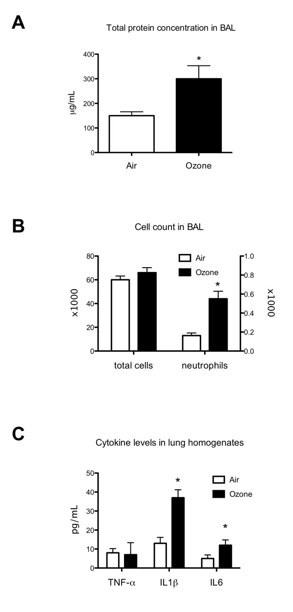
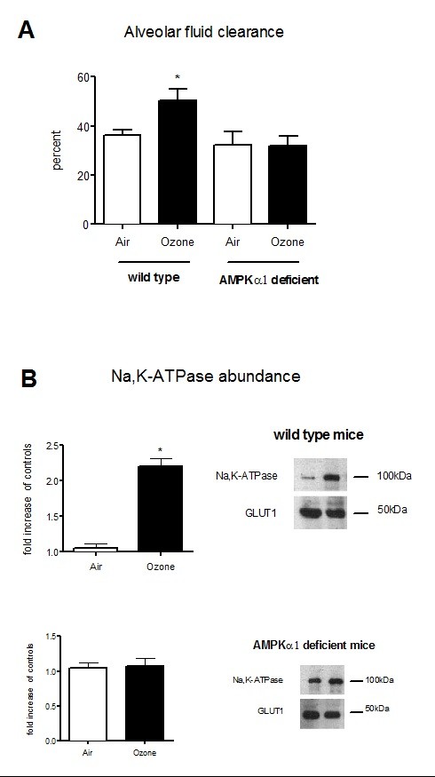
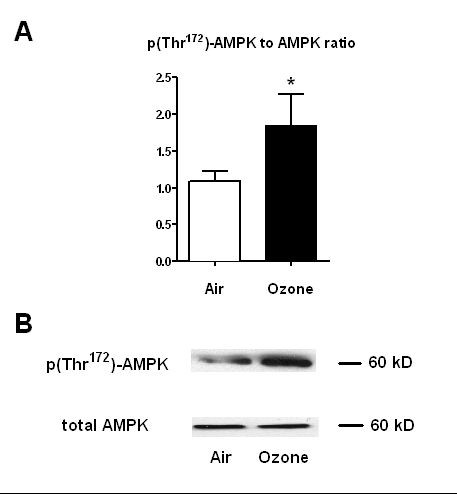
In control mice, ozone exposure induced lung inflammation as evidence by increased leukocyte count, protein concentration in BAL and myeloperoxidase activity, pro-inflammatory cytokine levels in LH. Increases in peroxynitrite levels (3 vs 4.4 nM, p = 0.02) and malondialdehyde concentrations (110 vs 230 μmole/g wet tissue) were detected in LH obtained from ozone-exposed control mice. Ozone exposure consistently increased phosphorylated AMPK-Thr172 to total AMPK ratio by 80% in control mice. Ozone exposure causes increases in AFC and basolateral membrane Na(+)-K(+)-ATPase abundance in control mice which did not occur in AMPKα1 deficient mice.
Our results collectively suggest that AMPK activation participates in ozone-induced increases in AFC, inflammation and oxidative stress. Further studies are needed to understand how the AMPK pathway may provide a novel approach for the prevention of ozone-induced lung injury.
急性臭氧暴露会导致肺部氧化应激和炎症,从而导致肺部损伤。臭氧肺毒性的至少一种机制涉及活性氧和氮中间产物(如过氧亚硝酸盐)的过度产生。此外,过氧亚硝酸盐可能会硝化酪氨酸残基,改变参与细胞信号转导的许多蛋白激酶的磷酸化,这超出了其主要的促氧化剂特性。最近有人提出,过氧亚硝酸盐会激活 5'-AMP 激活的蛋白激酶(AMPK),该激酶调节代谢途径和对细胞应激的反应。臭氧暴露后 AMPK 的激活尚未得到先前的评估。首先,我们测试了在小鼠中急性臭氧暴露是否会损害肺泡液清除率,增加肺组织过氧亚硝酸盐的产生并激活 AMPK。其次,我们测试了 AMPKα1 缺失是否会防止臭氧暴露引起的氧化应激增强和肺损伤。
在玻璃笼中,将对照和 AMPKα1 缺乏的小鼠暴露于 2.0 ppm 的臭氧中 3 小时。在臭氧暴露后 24 小时进行评估。使用异硫氰酸荧光素标记的白蛋白评估肺泡液清除率(AFC)。在支气管肺泡灌洗液(BAL)和肺匀浆(LH)中测定细胞计数、总蛋白水平、细胞因子浓度、髓过氧化物酶活性和氧化应激标志物(即丙二醛和过氧亚硝酸盐)。通过 Western blot 测定 AMPK-Thr172 磷酸化和基底外侧膜 Na(+)-K(+)-ATP 酶丰度。
在对照小鼠中,臭氧暴露引起肺部炎症,证据是白细胞计数、BAL 中的蛋白浓度和髓过氧化物酶活性增加,LH 中的促炎细胞因子水平升高。在对照小鼠的 LH 中检测到过氧亚硝酸盐水平(3 与 4.4 nM,p = 0.02)和丙二醛浓度(110 与 230 μmole/g 湿组织)增加。臭氧暴露使对照小鼠的磷酸化 AMPK-Thr172 与总 AMPK 的比值增加了 80%。臭氧暴露导致对照小鼠的 AFC 和基底外侧膜 Na(+)-K(+)-ATP 酶丰度增加,但在 AMPKα1 缺失小鼠中没有发生。
我们的结果表明,AMPK 激活参与了臭氧诱导的 AFC、炎症和氧化应激增加。需要进一步研究以了解 AMPK 途径如何为预防臭氧诱导的肺损伤提供新的方法。