Sofi Aijaz A, Thekdi Ashish D, Nawras Ali
Division of Gastroenterology, Department of Internal Medicine, University of Toledo Medical Center, 3000 Arlington Avenue, Toledo, OH 43614, USA.
Diagn Ther Endosc. 2011;2011:198029. doi: 10.1155/2011/198029. Epub 2011 Apr 6.
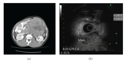
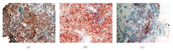
Primitive neuroectodermal tumor (PNET) is a rare "small round blue cell tumor" that is diagnosed by open biopsy or percutaneous biopsy of the lesion under radiologic guidance. In this case report, we present a novel approach to the diagnosis of a retroperitoneal PNET by endoscopic ultrasound- (EUS-) guided fine needle aspiration (FNA). A 35-year-old man presented with the history of left-sided flank pain and swelling of 3-weeks duration. Computerized tomography (CT) scan of his abdomen revealed a 12.8 × 13 × 12.5 cm cystic and solid mass arising from the retroperitoneum and displacing the third and fourth portions of the duodenum. He underwent EUS which revealed a well-circumscribed heterogeneous mass abutting the inferior portion of the stomach. EUS-FNA of the mass revealed malignant cells consistent with primitive neuroectodermal tumor (PNET)/Ewing's sarcoma. EUS-guided FNA is an appropriate technique for diagnosing retroperitoneal PNET/Ewing's sarcoma.
原始神经外胚层肿瘤(PNET)是一种罕见的“小圆蓝细胞瘤”,通过在放射学引导下对病变进行开放活检或经皮活检来诊断。在本病例报告中,我们展示了一种通过内镜超声(EUS)引导下细针穿刺抽吸(FNA)诊断腹膜后PNET的新方法。一名35岁男性,有左侧胁腹疼痛和肿胀3周的病史。他的腹部计算机断层扫描(CT)显示,一个12.8×13×12.5厘米的囊实性肿块起源于腹膜后,推移十二指肠第三和第四部分。他接受了EUS检查,发现一个边界清晰的异质性肿块紧邻胃下部。对该肿块进行EUS-FNA检查,发现恶性细胞,与原始神经外胚层肿瘤(PNET)/尤因肉瘤一致。EUS引导下FNA是诊断腹膜后PNET/尤因肉瘤的一种合适技术。