Shen Yi, Zhuang Pei, Chiou George C Y
Institute of Ocular Pharmacology, College of Medicine, Texas A&M Health Science Center, College Station, TX 77843, USA.
Open Ophthalmol J. 2011;5:27-31. doi: 10.2174/1874364101105010027. Epub 2011 Apr 28.
The effects of Guanabenz, an agonist of α2-adrenergic receptors routinely used in human medicine as an antihypertensive drug, were studied on NaIO(3)-induced retinal pigment epithelium (RPE) degeneration, laser-induced choroidal neovascularization (CNV) and choroidal blood flow, in animal models.
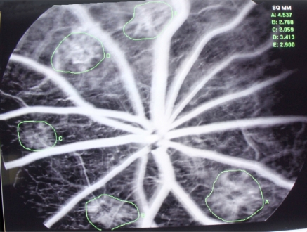
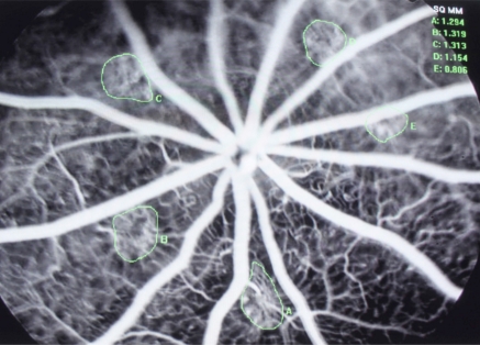
The 35mg/kg NaIO(3)-induced RPE degeneration rat eyes were instilled with 1% Guanabenz eye drops 3 times a day for 7 days before NaIO(3) injection, and then 2 to 4 weeks thereafter. RPE function was measured with c-wave of electroretinogram (ERG). Male Brown Norway rats were anesthetized to receive Nd:YAG laser to break the Bruch's membrane. One percent Guanabenz eye drops were given likewise. The development of CNV was determined by fluorescein angiography performed on week 2 and week 4 using sodium fluorescein (FA) or fluorescein isothiocyanatedextran (FD70-FA). Colored microsphere technique was used for in vivo experiments to determine the choroidal blood flow in ocular hypertensive (40 mmHg) rabbit eyes.
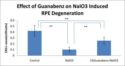
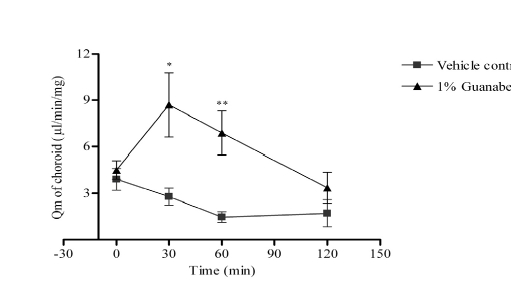
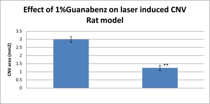
The RPE function was protected significantly by Guanabenz according to the c-wave of ERG. Four weeks after NaIO(3) injection, the amplitude of ERG c-wave was 0.422±0.092 millivolts in the control group, 0.103±0.04 millivolts in the NaIO(3) group, and 0.254±0.061 millivolts in the Guanabenz+NaIO(3) group. There was a significant protection of the ERG c-wave by Guanabenz as compared to NaIO(3) group (P<0.01). The angiograms of FD70-FA showed decreased lesion size in the Guanabenz group. Four weeks after laser treatment, the size of the CNV lesion was 2.99±0.18 mm(2) in the control group, and 1.24±0.16 mm(2) in the Guanabenz group (P<0.01). The choroidal blood flow was significantly increased at 30 and 60 minutes after Guanabenz instillation as compared to corresponding controls.
Guanabenz significantly protected RPE from NaIO(3)-induced degeneration, inhibited the development of CNV in laser-induced rat AMD model and increased choroidal blood flow markedly in vivo.
研究胍那苄(一种α2肾上腺素能受体激动剂,在人类医学中常作为抗高血压药物使用)对动物模型中碘酸钠(NaIO₃)诱导的视网膜色素上皮(RPE)变性、激光诱导的脉络膜新生血管(CNV)以及脉络膜血流的影响。
在注射NaIO₃前,对35mg/kg NaIO₃诱导RPE变性的大鼠眼睛每天滴注1%胍那苄滴眼液3次,持续7天,之后在2至4周内同样滴注。用电视网膜图(ERG)的c波测量RPE功能。对雄性棕色挪威大鼠进行麻醉,接受Nd:YAG激光以破坏布鲁赫膜。同样给予1%胍那苄滴眼液。在第2周和第周4使用荧光素钠(FA)或异硫氰酸荧光素葡聚糖(FD70 - FA)进行荧光素血管造影,以确定CNV的发展情况。采用彩色微球技术进行体内实验,以测定高眼压(40 mmHg)兔眼的脉络膜血流。
根据ERG的c波,胍那苄显著保护了RPE功能。注射NaIO₃后4周,对照组ERG c波振幅为0.422±0.092毫伏,NaIO₃组为0.103±0.04毫伏,胍那苄 + NaIO₃组为0.254±0.061毫伏。与NaIO₃组相比,胍那苄对ERG c波有显著保护作用(P<0.01)。FD70 - FA血管造影显示胍那苄组病变大小减小。激光治疗后4周,对照组CNV病变大小为2.99±0.18 mm²,胍那苄组为1.24±0.16 mm²(P<0.01)。与相应对照组相比,滴注胍那苄后30分钟和60分钟脉络膜血流显著增加。
胍那苄显著保护RPE免受NaIO₃诱导的变性,在激光诱导的大鼠年龄相关性黄斑变性(AMD)模型中抑制CNV的发展,并在体内显著增加脉络膜血流。