Endodontic Integrated Center, Passo Fundo, RS, Brazil.
J Appl Oral Sci. 2011 Aug;19(4):421-5. doi: 10.1590/s1678-77572011005000019. Epub 2011 Jul 1.
The objective of this work was to evaluate, using radiographic images, the behavior of four materials used to repair root perforations in dogs' teeth.
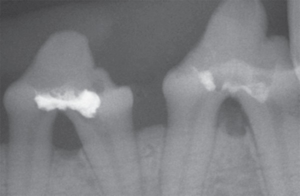
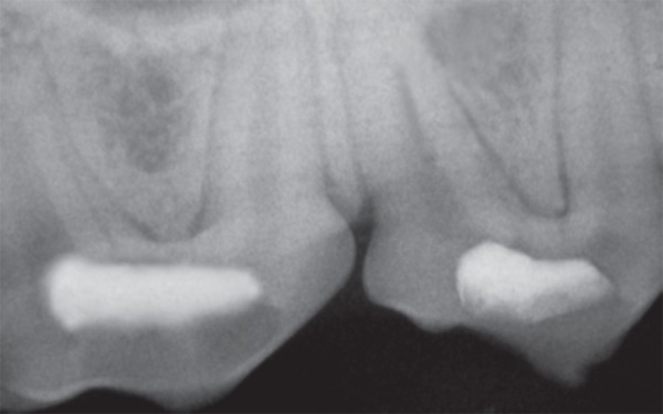
Second and third premolars of 6 dogs were used. The 48 teeth were randomly divided into 4 groups (n=12) and the perforations were sealed with one of the following materials: MTA, AH Plus, Vitremer and gutta-percha. Dogs were submitted to general anesthesia, teeth were radiographed and pulp was accessed. Perforations were done, at the maximum curve of the pulp floor, sealed and the accessed coronal cavity was filled with glass ionomer cement (Vidrion R). After 90 days, the dogs were sacrificed and the last x-ray image was taken. Images were analyzed for the presence/absence of periodontal lesions at the perforation region. Data were analyzed statistically by chi-square test at 5% significance level.
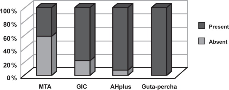
There were no statistically significant differences (p>0.05) among AH Plus, Vitremer and gutta-percha groups. MTA produced the smallest number of periodontal lesions (p<0.05).
It may be concluded that none of the tested materials was able to preserve the integrity of the periodontal tissues in the furcation region, and the use of MTA resulted in the least formation of adjacent periodontal bone lesions revealed by the radiographic comparisons.
本研究旨在通过影像学评估,研究 4 种材料在修复犬牙根管穿孔中的应用效果。
选取 6 只犬的第二和第三前磨牙,将 48 颗牙随机分为 4 组(n=12),分别使用 MTA、AH Plus、Vitremer 和牙胶进行根管穿孔修复。犬只接受全身麻醉,拍摄牙齿 X 光片,暴露牙髓腔。在牙髓腔底部最大弯曲处进行穿孔,用相应材料进行密封,然后用玻璃离子水门汀(Vidrion R)填充暴露的牙冠腔。90 天后,处死犬只,拍摄最后一张 X 光片。分析穿孔区域牙周病变的存在情况。采用卡方检验对数据进行统计学分析,检验水准为 5%。
AH Plus、Vitremer 和牙胶组之间无统计学差异(p>0.05)。MTA 组牙周病变数量最少(p<0.05)。
研究结果表明,测试的材料均不能维持牙周组织在分叉区的完整性,与其他材料相比,MTA 导致的相邻牙周骨病变最少。