• 文献检索
  • 文档翻译
  • 深度研究
  • 学术资讯
  • Suppr Zotero 插件Zotero 插件
  • 邀请有礼
  • 套餐&价格
  • 历史记录
应用&插件
Suppr Zotero 插件Zotero 插件浏览器插件Mac 客户端Windows 客户端微信小程序
定价
高级版会员购买积分包购买API积分包
服务
文献检索文档翻译深度研究API 文档MCP 服务
关于我们
关于 Suppr公司介绍联系我们用户协议隐私条款
关注我们

Suppr 超能文献

核心技术专利:CN118964589B侵权必究
粤ICP备2023148730 号-1Suppr @ 2026

文献检索

告别复杂PubMed语法,用中文像聊天一样搜索,搜遍4000万医学文献。AI智能推荐,让科研检索更轻松。

立即免费搜索

文件翻译

保留排版,准确专业,支持PDF/Word/PPT等文件格式,支持 12+语言互译。

免费翻译文档

深度研究

AI帮你快速写综述,25分钟生成高质量综述,智能提取关键信息,辅助科研写作。

立即免费体验

羊下颌骨牵张成骨和低水平激光治疗后硬度和弹性模量的初步研究。

A preliminary study of hardness and modulus of elasticity in sheep mandibles submitted to distraction osteogenesis and low-level laser therapy.

机构信息

School of Dentistry, Pontifícia Universidade Católica do Rio Grande do Sul (PUCRS), Porto Alegre, RS, Brazil.

出版信息

Med Oral Patol Oral Cir Bucal. 2012 Jan 1;17(1):e102-7. doi: 10.4317/medoral.17280.

DOI:10.4317/medoral.17280
PMID:21743401
原文链接:https://pmc.ncbi.nlm.nih.gov/articles/PMC3448184/
Abstract

OBJECTIVES

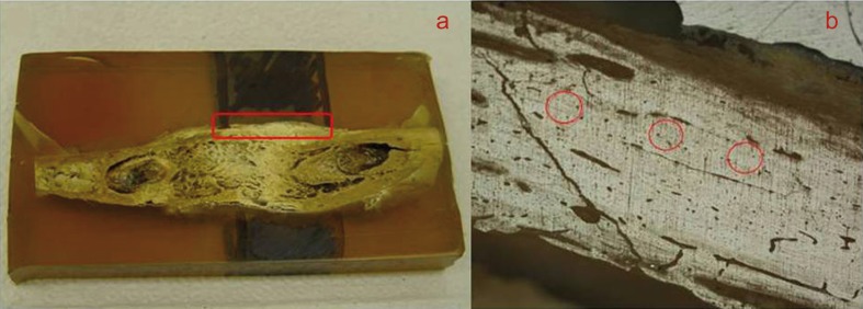
To investigate the quality of newly formed bone in sheep mandibles submitted to distraction osteogenesis and low-level laser therapy (LLLT), based on hardness and modulus of elasticity values. The ideal moment for laser application (during the latency/activation period vs. during the bone consolidation period) was also evaluated. Computed tomography imaging was used to assess relapse as a result of early device removal.

STUDY DESIGN

Extraoral distraction devices were placed in five sheep so as to achieve 1.5 cm of lengthened bone in 60 days. Distraction devices were removed 50, 40, and 33 days after surgery. Four animals were treated with LLLT, at different times, and one was used as control (no LLLT).

RESULTS

When applied during the bone consolidation period, LLLT caused an increase in hardness and modulus of elasticity values. On the other hand, animals irradiated with LLLT during the latency/activation period presented a delay in bone healing. A period of consolidation of 13 days (early device removal) was associated with relapse.

CONCLUSIONS

Nanoindentation tests were able to detect slight abnormalities in bone metabolism and proved to be important tools for the assessment of bone quality following distraction osteogenesis. LLLT provided increased benefits when applied during the bone consolidation period, once it promoted an increase in hardness and modulus of elasticity values. According to our results, the bone consolidation period should be of at least 3 weeks, so as to prevent relapse.

摘要

目的

通过硬度和弹性模量值,研究羊下颌骨接受牵张成骨和低水平激光治疗(LLLT)后新形成骨的质量。还评估了激光应用的理想时机(潜伏期/激活期与骨整合期)。计算机断层扫描成像用于评估因早期器械移除而导致的复发。

研究设计

在 5 只羊中放置体外牵张器,以在 60 天内实现 1.5 厘米的骨延长。手术后 50、40 和 33 天拆除牵张器。4 只动物接受了不同时间的 LLLT 治疗,1 只作为对照(未接受 LLLT)。

结果

LLLT 在骨整合期应用时,可增加硬度和弹性模量值。另一方面,在潜伏期/激活期接受 LLLT 照射的动物骨愈合延迟。13 天的整合期(早期器械移除)与复发有关。

结论

纳米压痕测试能够检测到骨代谢的轻微异常,是评估牵张成骨后骨质量的重要工具。LLLT 在骨整合期应用时提供了更大的益处,因为它可以提高硬度和弹性模量值。根据我们的结果,骨整合期至少应为 3 周,以防止复发。

https://cdn.ncbi.nlm.nih.gov/pmc/blobs/5998/3448184/189667bd3e51/medoral-17-e102-g003.jpg
https://cdn.ncbi.nlm.nih.gov/pmc/blobs/5998/3448184/fa7e7775c609/medoral-17-e102-g001.jpg
https://cdn.ncbi.nlm.nih.gov/pmc/blobs/5998/3448184/a56c821a51bc/medoral-17-e102-g002.jpg
https://cdn.ncbi.nlm.nih.gov/pmc/blobs/5998/3448184/189667bd3e51/medoral-17-e102-g003.jpg
https://cdn.ncbi.nlm.nih.gov/pmc/blobs/5998/3448184/fa7e7775c609/medoral-17-e102-g001.jpg
https://cdn.ncbi.nlm.nih.gov/pmc/blobs/5998/3448184/a56c821a51bc/medoral-17-e102-g002.jpg
https://cdn.ncbi.nlm.nih.gov/pmc/blobs/5998/3448184/189667bd3e51/medoral-17-e102-g003.jpg

相似文献

1
A preliminary study of hardness and modulus of elasticity in sheep mandibles submitted to distraction osteogenesis and low-level laser therapy.羊下颌骨牵张成骨和低水平激光治疗后硬度和弹性模量的初步研究。
Med Oral Patol Oral Cir Bucal. 2012 Jan 1;17(1):e102-7. doi: 10.4317/medoral.17280.
2
Finite element analysis of masticatory stress on neoformed bone tissue after distraction osteogenesis and low-level laser therapy: a pilot study.牵张成骨及低强度激光治疗后新生骨组织咀嚼应力的有限元分析:一项初步研究。
Photomed Laser Surg. 2014 Aug;32(8):429-36. doi: 10.1089/pho.2013.3671. Epub 2014 Jul 23.
3
Histological evaluation of the effect of low-level laser on distraction osteogenesis in rabbit mandibles.组织学评价低水平激光对兔下颌骨牵张成骨的影响。
Med Oral Patol Oral Cir Bucal. 2010 Jul 1;15(4):e616-8. doi: 10.4317/medoral.15.e616.
4
Evaluation of the effect of low-level diode laser therapy applied during the bone consolidation period following mandibular distraction osteogenesis in the human.评估低强度二极管激光疗法在人类下颌骨牵张成骨术后骨愈合期应用的效果。
Int J Oral Maxillofac Surg. 2015 Aug;44(8):989-97. doi: 10.1016/j.ijom.2015.04.010. Epub 2015 May 12.
5
Investigation of effects of low level laser therapy in distraction osteogenesis.研究低水平激光疗法在牵张成骨中的作用。
J Stomatol Oral Maxillofac Surg. 2018 Dec;119(6):469-476. doi: 10.1016/j.jormas.2018.05.006. Epub 2018 Jun 7.
6
Influence of a Magnetic Field and Laser Therapy on the Quality of Mandibular Bone During Distraction Osteogenesis in Rabbits.磁场和激光疗法对兔下颌骨牵张成骨过程中骨质量的影响
J Oral Maxillofac Surg. 2016 Nov;74(11):2287.e1-2287.e8. doi: 10.1016/j.joms.2016.07.010. Epub 2016 Jul 25.
7
A comparison of the low-level laser versus low intensity pulsed ultrasound on new bone formed through distraction osteogenesis.低强度激光与低强度脉冲超声对牵张成骨形成的新骨的比较。
Photomed Laser Surg. 2012 Aug;30(8):438-43. doi: 10.1089/pho.2012.3263. Epub 2012 Jul 10.
8
Effect of Laser Bio-Stimulation on Mandibular Distraction Osteogenesis: An Experimental Study.激光生物刺激对下颌骨牵张成骨的影响:一项实验研究。
J Oral Maxillofac Surg. 2018 Nov;76(11):2411-2421. doi: 10.1016/j.joms.2018.04.030. Epub 2018 May 7.
9
Bone tissue microscopic findings related to the use of diode laser (830 nm) in ovine mandible submitted to distraction osteogenesis.与在接受牵张成骨的绵羊下颌骨中使用二极管激光(830纳米)相关的骨组织显微镜检查结果。
Acta Cir Bras. 2007 Mar-Apr;22(2):92-7. doi: 10.1590/s0102-86502007000200003.
10
Effects of low-level laser therapy on bone formed after distraction osteogenesis.低水平激光治疗对牵张成骨后形成的骨的影响。
Lasers Med Sci. 2010 Mar;25(2):213-9. doi: 10.1007/s10103-009-0691-2. Epub 2009 Jun 23.

引用本文的文献

1
External devices increasing bone quality in animals: A systematic review.增加动物骨质量的外部装置:一项系统综述。
Heliyon. 2023 Nov 16;9(11):e22379. doi: 10.1016/j.heliyon.2023.e22379. eCollection 2023 Nov.
2
Overview of Physical and Pharmacological Therapy in Enhancing Bone Regeneration Formation During Distraction Osteogenesis.牵引成骨过程中促进骨再生形成的物理和药物治疗概述
Front Cell Dev Biol. 2022 Apr 28;10:837430. doi: 10.3389/fcell.2022.837430. eCollection 2022.
3
The Effect of Photobiomodulation on Distraction Osteogenesis.

本文引用的文献

1
Histological evaluation of the effect of low-level laser on distraction osteogenesis in rabbit mandibles.组织学评价低水平激光对兔下颌骨牵张成骨的影响。
Med Oral Patol Oral Cir Bucal. 2010 Jul 1;15(4):e616-8. doi: 10.4317/medoral.15.e616.
2
Analysis of the systemic effect of red and infrared laser therapy on wound repair.红激光与红外激光疗法对伤口愈合的全身效应分析
Photomed Laser Surg. 2009 Dec;27(6):929-35. doi: 10.1089/pho.2008.2306.
3
Effects of low-level laser therapy on bone formed after distraction osteogenesis.低水平激光治疗对牵张成骨后形成的骨的影响。
光生物调节对牵张成骨的影响。
J Lasers Med Sci. 2019 Fall;10(4):330-337. doi: 10.15171/jlms.2019.53. Epub 2019 Oct 1.
4
The assessment of new bone formation induced by unfocused extracorporeal shock wave therapy applied on pre-surgical phase of distraction osteogenesis.在牵张成骨术前阶段应用非聚焦体外冲击波疗法诱导新骨形成的评估。
Eur Oral Res. 2019 Sep;53(3):125-131. doi: 10.26650/eor.20190041. Epub 2019 Sep 1.
5
Systemic effects of LLLT on bone repair around PLLA-PGA screws in the rabbit tibia.
Lasers Med Sci. 2014 Mar;29(2):703-8. doi: 10.1007/s10103-013-1384-4. Epub 2013 Jul 6.
Lasers Med Sci. 2010 Mar;25(2):213-9. doi: 10.1007/s10103-009-0691-2. Epub 2009 Jun 23.
4
Effect of low-level laser therapy after implantation of poly-L-lactic/polyglycolic acid in the femurs of rats.
Lasers Med Sci. 2009 Sep;24(5):721-8. doi: 10.1007/s10103-008-0627-2. Epub 2008 Nov 15.
5
Changes in blood perfusion and bone healing induced by nicotine during distraction osteogenesis.尼古丁在牵张成骨过程中引起的血液灌注变化和骨愈合
Bone. 2008 Aug;43(2):355-361. doi: 10.1016/j.bone.2008.04.002. Epub 2008 Apr 16.
6
The influence of expansion rates on mandibular distraction osteogenesis: a computational analysis.扩张速率对下颌骨牵张成骨的影响:一项计算分析。
Ann Biomed Eng. 2007 Nov;35(11):1940-60. doi: 10.1007/s10439-007-9367-x. Epub 2007 Sep 4.
7
Bone tissue microscopic findings related to the use of diode laser (830 nm) in ovine mandible submitted to distraction osteogenesis.与在接受牵张成骨的绵羊下颌骨中使用二极管激光(830纳米)相关的骨组织显微镜检查结果。
Acta Cir Bras. 2007 Mar-Apr;22(2):92-7. doi: 10.1590/s0102-86502007000200003.
8
Low-level laser effect on mandibular distraction osteogenesis.低强度激光对下颌骨牵张成骨的影响。
J Oral Maxillofac Surg. 2007 Feb;65(2):168-76. doi: 10.1016/j.joms.2006.10.002.
9
Distraction osteogenesis of the porcine mandible: histomorphometric evaluation of bone.猪下颌骨的牵张成骨:骨组织形态计量学评估
Plast Reconstr Surg. 2004 Feb;113(2):566-73. doi: 10.1097/01.PRS.0000101061.99577.09.
10
Does the sheep mandible relapse following lengthening by distraction osteogenesis?绵羊下颌骨经牵张成骨延长后会复发吗?
J Craniomaxillofac Surg. 2000 Oct;28(5):251-7. doi: 10.1054/jcms.2000.0153.