Department of Anesthesiology, Ohio State University, Columbus, Ohio.
Inflamm Bowel Dis. 2011 Aug;17(8):1698-713. doi: 10.1002/ibd.21553. Epub 2010 Dec 3.
Pharmacological studies suggest that adenosine A₃AR influences motility and colitis. Functional A₃⁻/⁻AR knockout mice were used to prove whether A₃AR activation is involved in modulating either motility or colitis.
A₃AR was probed by polymerase chain reaction (PCR) genotyping, Western blot, and immunochemistry. Motility was assessed in vivo by artificial bead-expulsion, stool-frequency, and FITC-dextran transit. Colitis was induced with dextran sodium sulfate (DSS) in A₃⁻/⁻AR or wildtype (WT) age- and sex-matched controls. Progression of colitis was evaluated by histopathology, changes in myeloperoxidase (MPO), colon length, CD4(+) -cells, weight-loss, diarrhea, and the guaiac test.
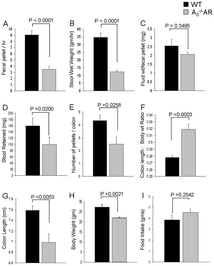
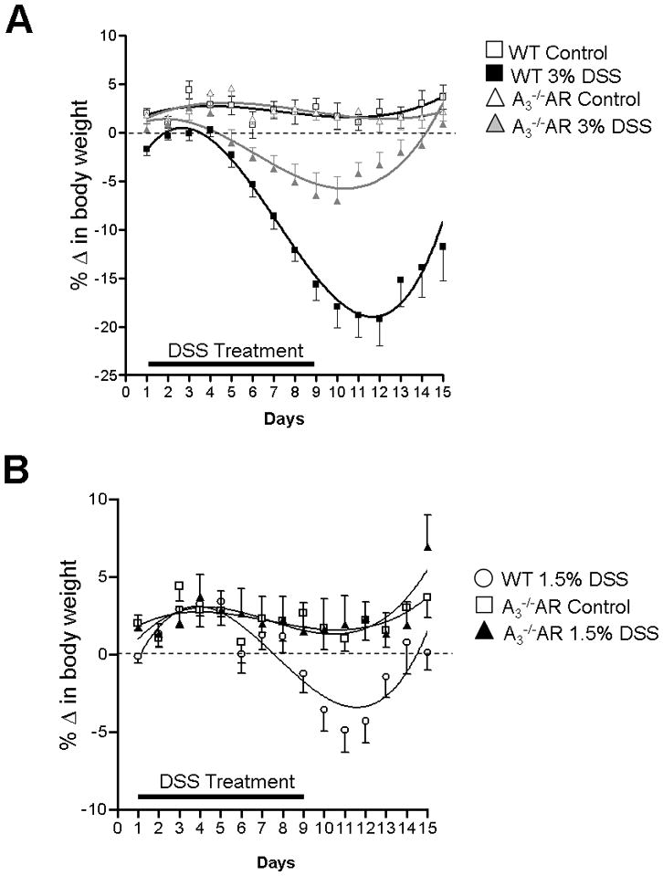
Goat anti-hu-A₃ antiserum identified a 66 kDa immunogenic band in colon. A₃AR-immunoreactivity is expressed in SYN(+) -nerve varicosities, s-100(+) -glia, and crypt cells, but not 5-HT(+) (EC), CD4(+) (T), tryptase(+) (MC), or muscle cells. A₃AR immunoreactivity in myenteric ganglia of distal colon >> proximal colon by a ratio of 2:1. Intestinal transit and bead expulsion were accelerated in A₃⁻/⁻AR mice compared to WT; stool retention was lower by 40%-60% and stool frequency by 67%. DSS downregulated A₃AR in epithelia. DSS histopathology scores indicated less mucosal damage in AA₃⁻/⁻AR mice than WT. A₃⁻/⁻AR phenotype protected against DSS-induced weight loss, neutrophil (MPO), or CD4(+) -T cell infiltration, colon shortening, change in splenic weight, diarrhea, or occult-fecal blood.
Functional disruption of A₃AR in A₃⁻/⁻AR mice alters intestinal motility. We postulate that ongoing release of adenosine and activation of presynaptic-inhibitory A₃AR can slow down transit and inhibit the defecation reflex. A₃AR may be involved in gliotransmission. In separate studies, A₃⁻/⁻AR protects against DSS colitis, consistent with a novel hypothesis that A₃AR activation contributes to development of colitis.
药理学研究表明,腺苷 A₃AR 会影响运动和结肠炎。使用功能性 A₃⁻/⁻AR 基因敲除小鼠来证明 A₃AR 的激活是否参与调节运动或结肠炎。
通过聚合酶链反应 (PCR) 基因分型、Western blot 和免疫化学方法探测 A₃AR。通过人工珠排出、粪便频率和 FITC-葡聚糖转运来体内评估运动。在 A₃⁻/⁻AR 或野生型 (WT) 年龄和性别匹配的对照中,用葡聚糖硫酸钠 (DSS) 诱导结肠炎。通过组织病理学、髓过氧化物酶 (MPO) 变化、结肠长度、CD4(+) -细胞、体重减轻、腹泻和愈创木脂测试评估结肠炎的进展。
山羊抗 hu-A₃ 抗血清在结肠中识别出 66 kDa 的免疫原性带。A₃AR-免疫反应性表达在 SYN(+) -神经轴突、s-100(+) -胶质细胞和隐窝细胞中,但不在 5-HT(+) (EC)、CD4(+) (T)、胰蛋白酶(+) (MC) 或肌肉细胞中。远端结肠的 A₃AR 免疫反应性比近端结肠高 2:1。与 WT 相比,A₃⁻/⁻AR 小鼠的肠道转运和珠排出加快;粪便保留率降低 40%-60%,粪便频率增加 67%。DSS 在肠上皮下调 A₃AR。DSS 组织病理学评分表明,与 WT 相比,AA₃⁻/⁻AR 小鼠的黏膜损伤更小。A₃⁻/⁻AR 表型可预防 DSS 诱导的体重减轻、中性粒细胞 (MPO) 或 CD4(+) -T 细胞浸润、结肠缩短、脾重变化、腹泻或隐血粪便。
在 A₃⁻/⁻AR 小鼠中,A₃AR 的功能破坏改变了肠道运动。我们推测,持续释放的腺苷和激活的突触前抑制性 A₃AR 可以减缓转运并抑制排便反射。A₃AR 可能参与神经胶质传递。在单独的研究中,A₃⁻/⁻AR 可预防 DSS 结肠炎,这与一种新的假说一致,即 A₃AR 的激活有助于结肠炎的发展。