Division of Signal Transduction and Growth Control (A100), DKFZ-ZMBH Alliance, German Cancer Research Center, Heidelberg, Germany.
J Invest Dermatol. 2011 Nov;131(11):2281-8. doi: 10.1038/jid.2011.190. Epub 2011 Jul 14.
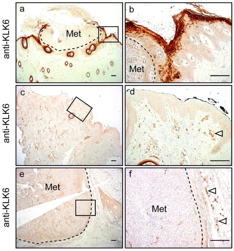
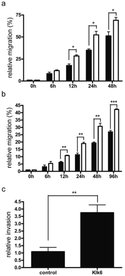
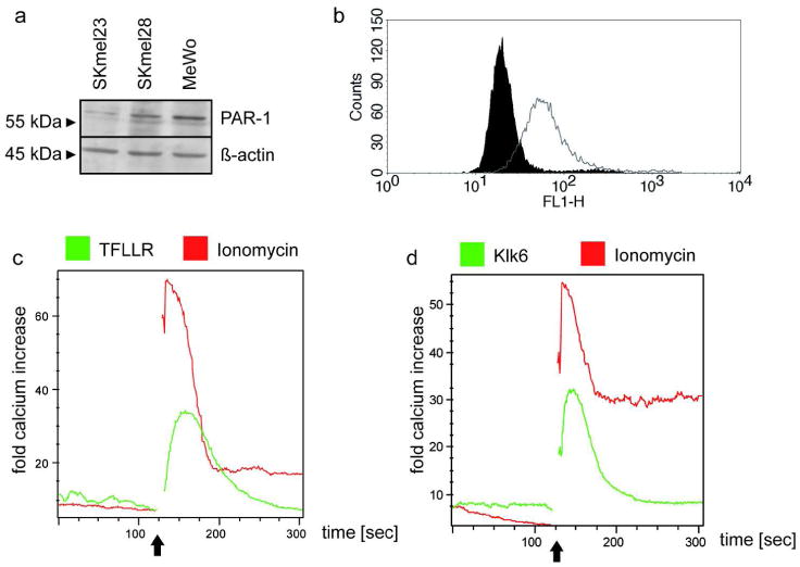
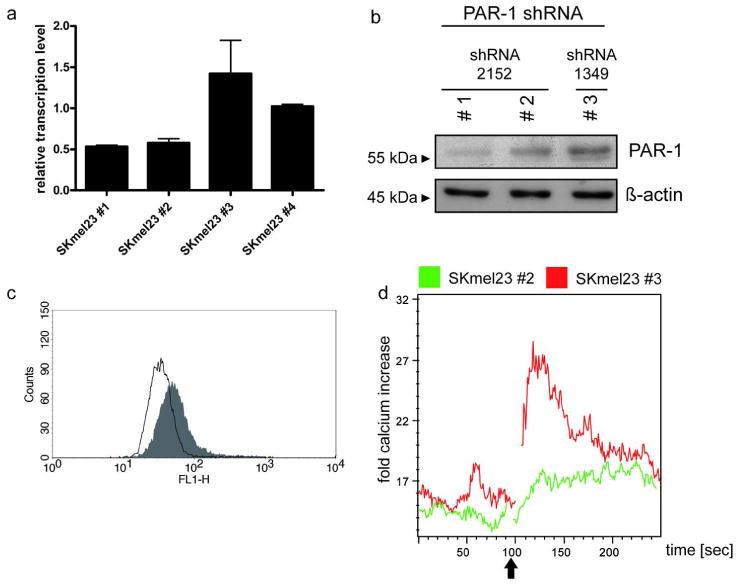
Cutaneous malignant melanoma is an aggressive disease of poor prognosis. Clinical and experimental studies have provided major insight into the pathogenesis of the disease, including the functional interaction between melanoma cells and surrounding keratinocytes, fibroblasts, and immune cells. Nevertheless, patients with metastasized melanoma have a very poor prognosis and are largely refractory to clinical therapies. Hence, diagnostic tools to monitor melanoma development, as well as therapeutic targets, are urgently needed. We investigated the expression pattern of the kallikrein-related peptidase 6 (KLK6) in human melanoma tissue sections throughout tumor development. Although KLK6 was not detectable in tumor cells, we found strong KLK6 protein expression in keratinocytes and stromal cells located adjacent to benign nevi, primary melanomas, and cutaneous metastatic lesions, suggesting a paracrine function of extracellular KLK6 during neoplastic transformation and malignant progression. Accordingly, recombinant Klk6 protein significantly induced melanoma cell migration and invasion accompanied by an accelerated intracellular Ca(2+) flux. We could further demonstrate that KLK6-induced intracellular Ca(2+) flux and tumor cell invasion critically depends on the protease-activated receptor 1 (PAR1). Our data provide experimental evidence that specific inhibition of the KLK6-PAR1 axis may interfere with the deleterious effect of tumor-microenvironment interaction and represent a potential option for translational melanoma research.
皮肤恶性黑色素瘤是一种预后不良的侵袭性疾病。临床和实验研究为该疾病的发病机制提供了重要的认识,包括黑色素瘤细胞与周围角质形成细胞、成纤维细胞和免疫细胞之间的功能相互作用。然而,转移性黑色素瘤患者的预后非常差,并且对临床治疗大多具有抗性。因此,迫切需要用于监测黑色素瘤发展的诊断工具以及治疗靶标。我们研究了激肽释放酶相关肽酶 6(KLK6)在人类黑色素瘤组织切片中的表达模式,这些组织切片涵盖了肿瘤发展的全过程。尽管在肿瘤细胞中无法检测到 KLK6,但我们在位于良性痣、原发性黑色素瘤和皮肤转移性病变附近的角质形成细胞和成纤维细胞中发现了强烈的 KLK6 蛋白表达,这表明细胞外 KLK6 在肿瘤转化和恶性进展过程中具有旁分泌功能。相应地,重组 Klk6 蛋白显著诱导黑色素瘤细胞迁移和侵袭,同时伴随着细胞内 Ca(2+)流的加速。我们还进一步证明 KLK6 诱导的细胞内 Ca(2+)流和肿瘤细胞侵袭严重依赖蛋白酶激活受体 1(PAR1)。我们的数据提供了实验证据,表明 KLK6-PAR1 轴的特异性抑制可能会干扰肿瘤微环境相互作用的有害影响,这代表了转化性黑色素瘤研究的一个潜在选择。