Department of Molecular Medicine, Mayo Clinic, Rochester, MN 55905, USA.
Gene Ther. 2012 Mar;19(3):279-87. doi: 10.1038/gt.2011.107. Epub 2011 Jul 14.
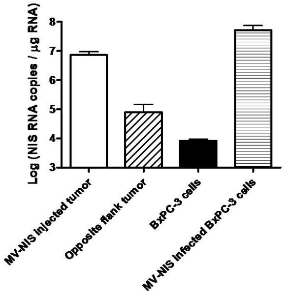
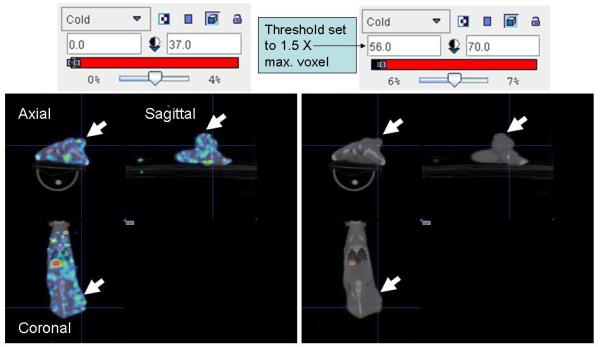
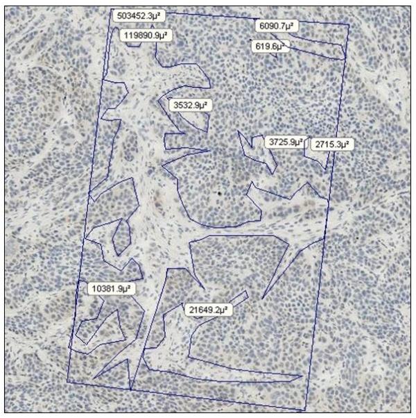
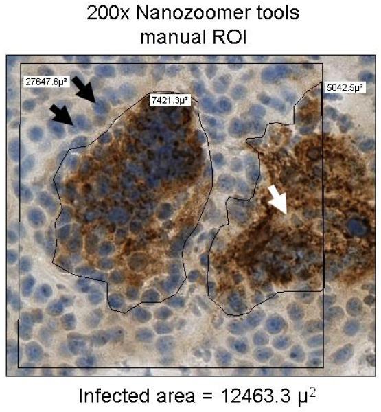
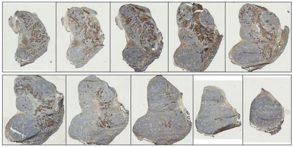
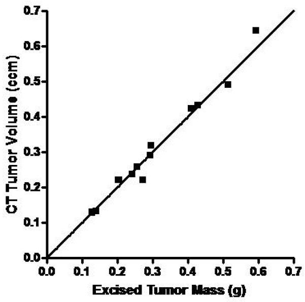
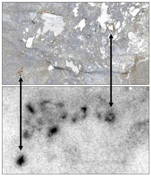
The purpose of our study was to validate the ability of pinhole micro-single-photon emission computed tomography/computed tomography (SPECT/CT) to: 1) accurately resolve the intratumoral dispersion pattern and 2) quantify the infection percentage in solid tumors of an oncolytic measles virus encoding the human sodium iodide symporter (MV-NIS). Sodium iodide symporter (NIS) RNA level and dispersion pattern were determined in control and MV-NIS-infected BxPC-3 pancreatic tumor cells and mouse xenografts using quantitative, real-time, reverse transcriptase, polymerase chain reaction, autoradiography and immunohistochemistry (IHC). Mice with BxPC-3 xenografts were imaged with (123)I or (99)TcO(4) micro-SPECT/CT. Tumor dimensions and radionuclide localization were determined with imaging software. Linear regression and correlation analyses were performed to determine the relationship between tumor infection percentage and radionuclide uptake (% injected dose per gram) above background and a highly significant correlation was observed (r(2)=0.947). A detection threshold of 1.5-fold above the control tumor uptake (background) yielded a sensitivity of 2.7% MV-NIS-infected tumor cells. We reliably resolved multiple distinct intratumoral zones of infection from non-infected regions. Pinhole micro-SPECT/CT imaging using the NIS reporter demonstrated precise localization and quantitation of oncolytic MV-NIS infection, and can replace more time-consuming and expensive analyses (for example, autoradiography and IHC) that require animal killing.
我们研究的目的是验证微孔单光子发射计算机断层扫描/计算机断层扫描(SPECT/CT)的能力:1)准确分辨肿瘤内的分散模式,2)定量测量溶瘤麻疹病毒编码人钠碘转运体(MV-NIS)的实体瘤中的感染百分比。采用定量、实时逆转录聚合酶链反应、放射性自显影和免疫组织化学(IHC)方法,在对照和 MV-NIS 感染的 BxPC-3 胰腺肿瘤细胞和小鼠异种移植瘤中测定钠碘转运体(NIS)RNA 水平和分散模式。用(123)I 或(99)TcO(4)微孔 SPECT/CT 对携带 BxPC-3 异种移植瘤的小鼠进行成像。用成像软件确定肿瘤的尺寸和放射性核素定位。进行线性回归和相关性分析,以确定肿瘤感染百分比与放射性核素摄取(每克注射剂量的百分比)与背景的关系,观察到高度显著的相关性(r(2)=0.947)。与对照肿瘤摄取(背景)相比,检测阈值高出 1.5 倍,可检测到 2.7%的 MV-NIS 感染肿瘤细胞。我们可靠地从非感染区域分辨出多个不同的肿瘤内感染区域。使用 NIS 报告基因的微孔 SPECT/CT 成像能够精确定位和定量溶瘤 MV-NIS 感染,并且可以替代更耗时且昂贵的分析(例如放射性自显影和 IHC),这些分析需要杀死动物。