Bal Cenkhan, Alacam Alev, Tuzuner Tamer, Tirali Resmiye Ebru, Baris Emre
Department of Paediatric Dentistry, Faculty of Dentistry, Gazi University, Ankara, Turkey.
Eur J Dent. 2011 Jul;5(3):265-72.
The objective of this pilot study was to evaluate the effects of three different antiseptic materials on healing processes of direct pulp therapies with Ca(OH)(2) histopathologically.
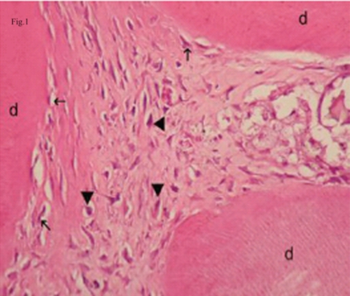
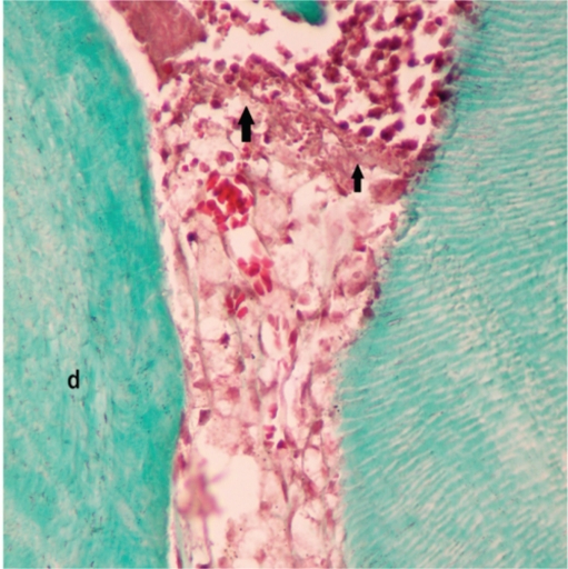
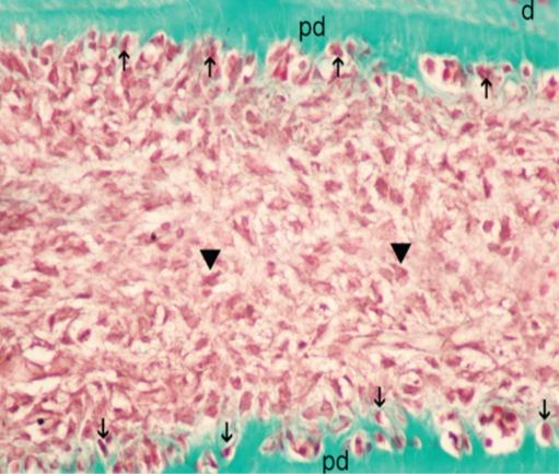
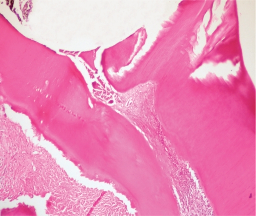
Twenty-eight upper and lower first molar teeth from 7 male Wistar rats were used in this study. Four cavities were prepared in each rat in four quadrants, and each quadrant represented different experimental groups. In Group I: 0.5% sodium hypochlorite (NaOCl); in Group II: 2% chlorhexidine digluconate (CHX); in Group III: 0.1% octenidine dihydrochloride (OCT); and in Group IV 0.9% sterile saline was applied to the exposure site with a sterile cotton pellet for 3 minutes. After hemorrhage control, the pulps were capped with hard setting Ca(OH)(2) and, finally, restored with IRM. The animals were euthanized at 21 days post-operatively. After sacrificing, routine histological procedures were performed and evaluated statistically with non-parametric Kruskal-Wallis test among the groups and two-by-two comparisons by using the Mann-Whitney U test for inflammatory response and tissue organization scores at the confidence interval of 95%.
There were significant differences in inflammatory response and tissue organization scores between the groups (P<.05). Statistical evaluation of inflammatory response showed that Group IV was significantly different from Groups I, II and III separately with a higher inflammatory cell response (P<.05) whereas no significant differences were detected between the other groups in two-by-two comparisons (P>.05). Healthy coronal and radicular pulp tissue organization scores indicated that the Group I has better pulp tissue organization than Group IV and this was significantly different (P<.05) whereas no significant differences were observed between the other groups separately (P>.05).
The antiseptic materials used in this study created an environment that, rather than saline solution, may affect clinical and histological success in a positive way.
本初步研究的目的是从组织病理学角度评估三种不同抗菌材料对用氢氧化钙进行直接牙髓治疗愈合过程的影响。
本研究使用了7只雄性Wistar大鼠的28颗上下颌第一磨牙。在每只大鼠的四个象限各制备四个洞,每个象限代表不同的实验组。第一组:0.5%次氯酸钠(NaOCl);第二组:2%葡萄糖酸洗必泰(CHX);第三组:0.1%二盐酸奥替尼啶(OCT);第四组用无菌棉球蘸取0.9%无菌生理盐水涂抹于暴露部位3分钟。控制出血后,用硬固型氢氧化钙覆盖牙髓,最后用IRM进行修复。术后21天对动物实施安乐死。处死后,进行常规组织学程序,并在各组间进行非参数Kruskal-Wallis检验,并使用Mann-Whitney U检验在95%置信区间对炎症反应和组织学结构评分进行两两比较,进行统计学评估。
各组间炎症反应和组织学结构评分存在显著差异(P<0.05)。炎症反应的统计学评估显示,第四组与第一、二、三组分别有显著差异,炎症细胞反应更高(P<0.05),而其他组两两比较未检测到显著差异(P>0.05)。健康的冠部和根部牙髓组织结构评分表明,第一组比第四组有更好的牙髓组织结构,且差异显著(P<0.05),而其他组两两比较未观察到显著差异(P>0.05)。
本研究中使用的抗菌材料创造了一种环境,与生理盐水相比,可能以积极的方式影响临床和组织学成功率。