Liver Imaging Group, Department of Radiology, University of California, San Diego Medical Center, University of California at San Diego, MR3T Laboratory, San Diego, California 92103-8226, USA.
J Magn Reson Imaging. 2011 Oct;34(4):928-34. doi: 10.1002/jmri.22701. Epub 2011 Jul 18.
To evaluate magnetic resonance imaging (MRI)-determined proton density fat fraction (PDFF) reproducibility across two MR scanner platforms and, using MR spectroscopy (MRS)-determined PDFF as reference standard, to confirm MRI-determined PDFF estimation accuracy.
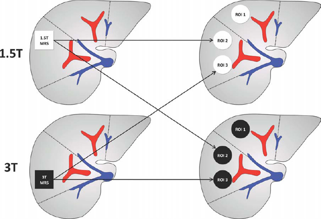
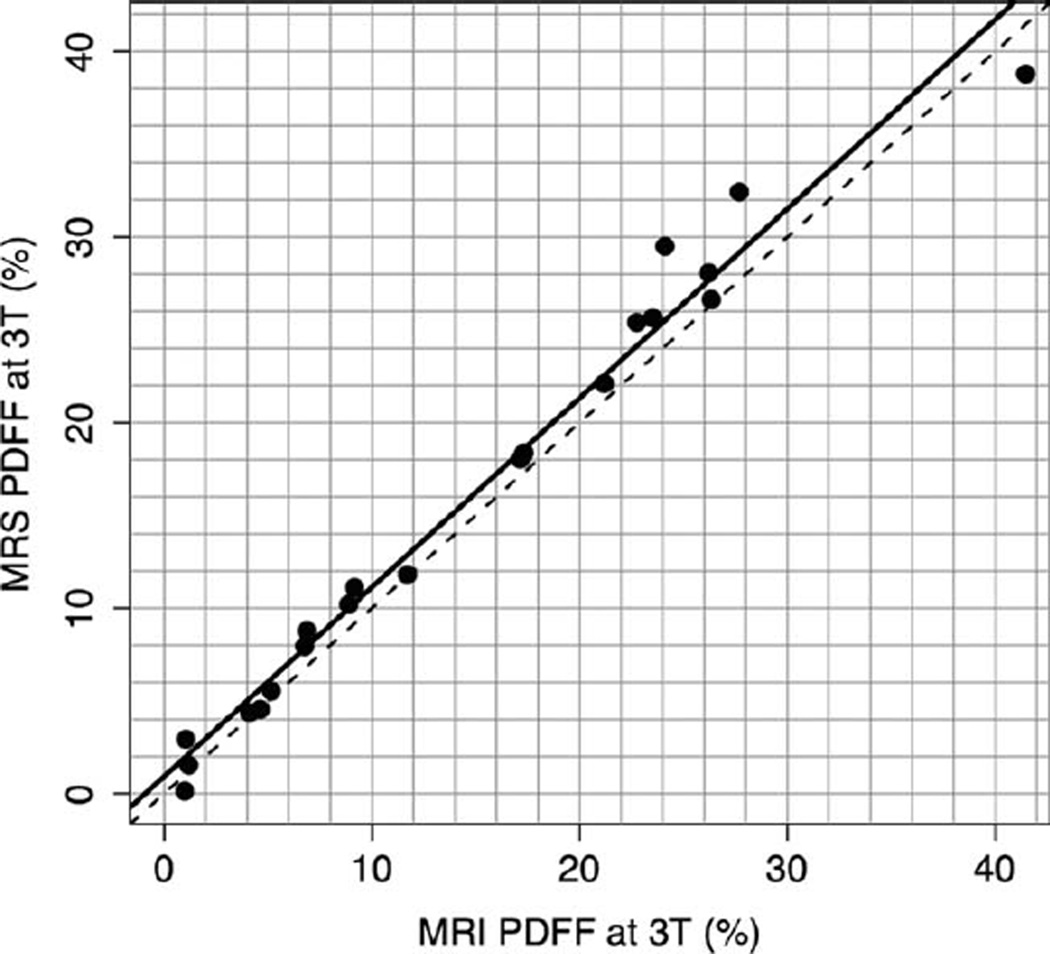
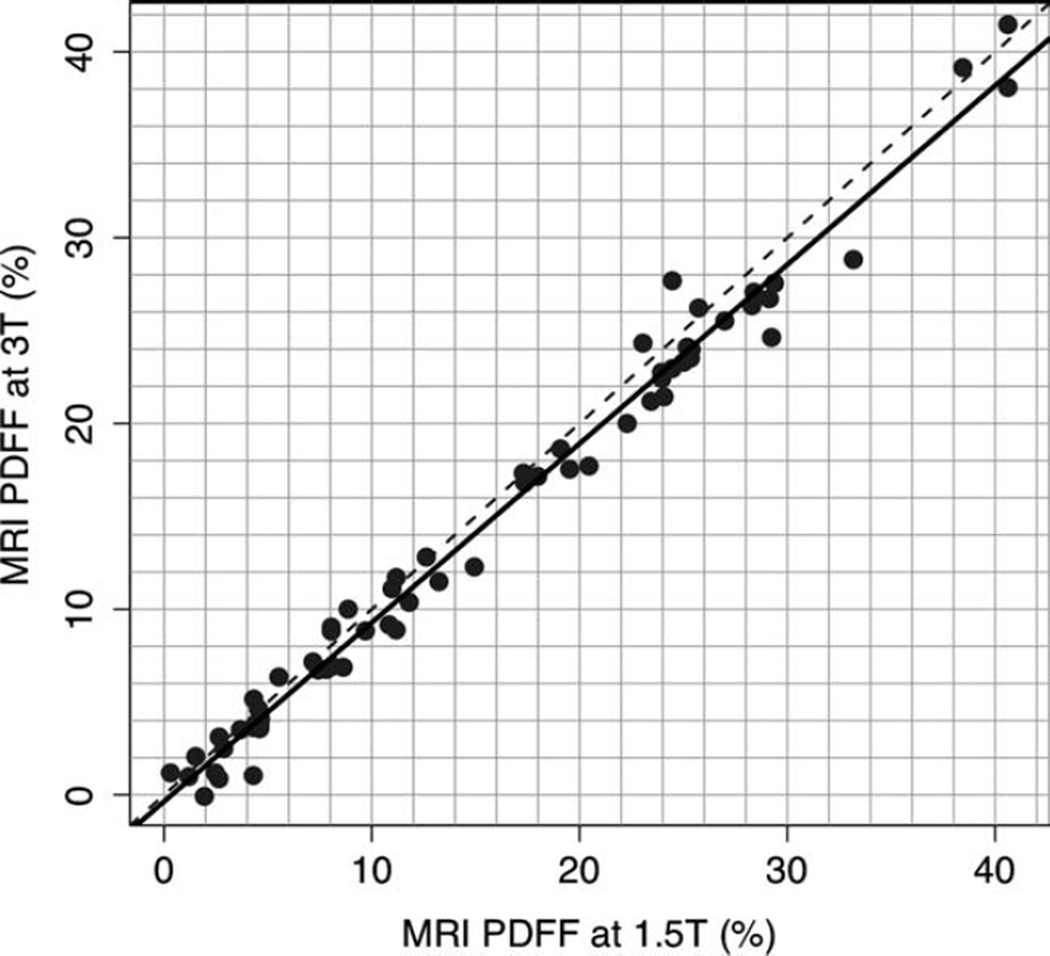
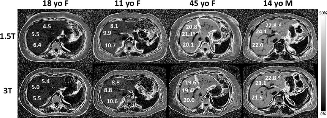
This prospective, cross-sectional, crossover, observational pilot study was approved by an Institutional Review Board. Twenty-one subjects gave written informed consent and underwent liver MRI and MRS at both 1.5T (Siemens Symphony scanner) and 3T (GE Signa Excite HD scanner). MRI-determined PDFF was estimated using an axial 2D spoiled gradient-recalled echo sequence with low flip-angle to minimize T1 bias and six echo-times to permit correction of T2* and fat-water signal interference effects. MRS-determined PDFF was estimated using a stimulated-echo acquisition mode sequence with long repetition time to minimize T1 bias and five echo times to permit T2 correction. Interscanner reproducibility of MRI determined PDFF was assessed by correlation analysis; accuracy was assessed separately at each field strength by linear regression analysis using MRS-determined PDFF as reference standard.
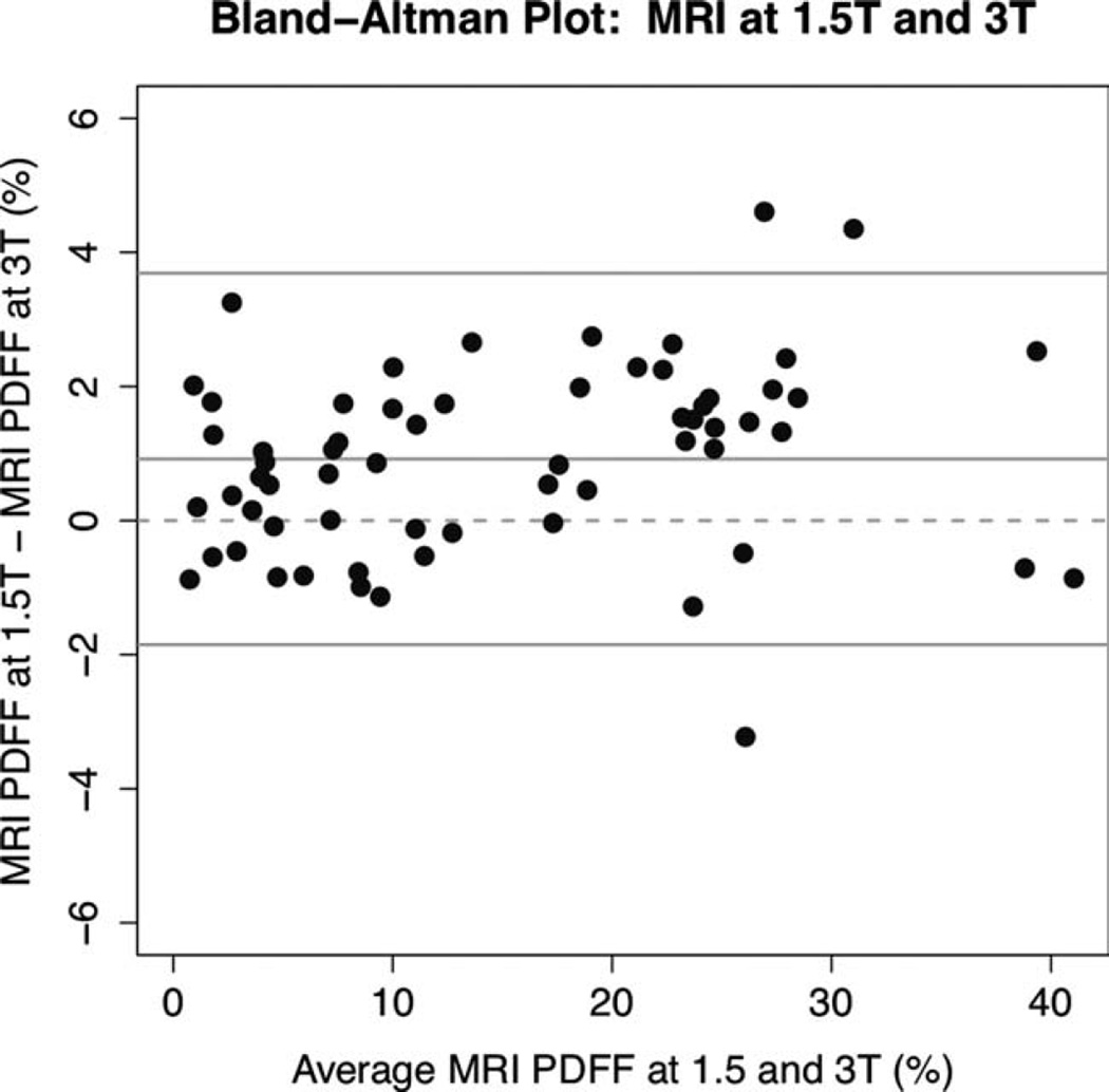
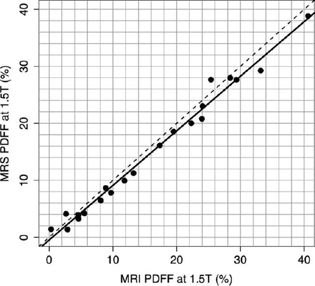
1.5T and 3T MRI-determined PDFF estimates were highly correlated (r = 0.992). MRI-determined PDFF estimates were accurate at both 1.5T (regression slope/intercept = 0.958/-0.48) and 3T (slope/intercept = 1.020/0.925) against the MRS-determined PDFF reference.
MRI-determined PDFF estimation is reproducible and, using MRS-determined PDFF as reference standard, accurate across two MR scanner platforms at 1.5T and 3T.
评估两种磁共振扫描仪平台上磁共振成像(MRI)确定的质子密度脂肪分数(PDFF)的可重复性,并使用磁共振波谱(MRS)确定的 PDFF 作为参考标准,确认 MRI 确定的 PDFF 估计的准确性。
本前瞻性、横断面、交叉、观察性初步研究获得了机构审查委员会的批准。21 名受试者签署了书面知情同意书,并在 1.5T(西门子 Symphony 扫描仪)和 3T(GE Signa Excite HD 扫描仪)上进行了肝脏 MRI 和 MRS 检查。使用具有低翻转角的轴向 2D 扰相梯度回波序列估计 MRI 确定的 PDFF,以最小化 T1 偏差和六个回波时间,以允许校正 T2*和脂肪-水信号干扰效应。使用具有长重复时间的受激回波采集模式序列估计 MRS 确定的 PDFF,以最小化 T1 偏差和五个回波时间,以允许 T2 校正。通过相关分析评估 MRI 确定的 PDFF 的扫描仪间可重复性;分别在每个场强下通过线性回归分析评估准确性,以 MRS 确定的 PDFF 作为参考标准。
1.5T 和 3T MRI 确定的 PDFF 估计值高度相关(r = 0.992)。MRI 确定的 PDFF 估计值在 1.5T(回归斜率/截距= 0.958/-0.48)和 3T(斜率/截距= 1.020/0.925)处均与 MRS 确定的 PDFF 参考值准确。
MRI 确定的 PDFF 估计具有可重复性,并且使用 MRS 确定的 PDFF 作为参考标准,在 1.5T 和 3T 两种磁共振扫描仪平台上均准确。