Translational Cytomics Group, Department of Surgery, Cedars-Sinai Medical Center, Los Angeles, California, United States of America.
PLoS One. 2011;6(7):e21861. doi: 10.1371/journal.pone.0021861. Epub 2011 Jul 14.
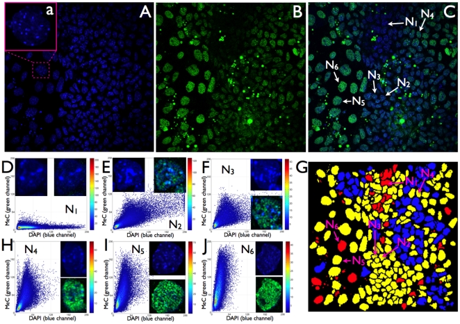
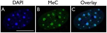
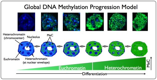
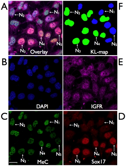
The genome organization in pluripotent cells undergoing the first steps of differentiation is highly relevant to the reprogramming process in differentiation. Considering this fact, chromatin texture patterns that identify cells at the very early stage of lineage commitment could serve as valuable tools in the selection of optimal cell phenotypes for regenerative medicine applications. Here we report on the first-time use of high-resolution three-dimensional fluorescence imaging and comprehensive topological cell-by-cell analyses with a novel image-cytometrical approach towards the identification of in situ global nuclear DNA methylation patterns in early endodermal differentiation of mouse ES cells (up to day 6), and the correlations of these patterns with a set of putative markers for pluripotency and endodermal commitment, and the epithelial and mesenchymal character of cells. Utilizing this in vitro cell system as a model for assessing the relationship between differentiation and nuclear DNA methylation patterns, we found that differentiating cell populations display an increasing number of cells with a gain in DNA methylation load: first within their euchromatin, then extending into heterochromatic areas of the nucleus, which also results in significant changes of methylcytosine/global DNA codistribution patterns. We were also able to co-visualize and quantify the concomitant stochastic marker expression on a per-cell basis, for which we did not measure any correlation to methylcytosine loads or distribution patterns. We observe that the progression of global DNA methylation is not correlated with the standard transcription factors associated with endodermal development. Further studies are needed to determine whether the progression of global methylation could represent a useful signature of cellular differentiation. This concept of tracking epigenetic progression may prove useful in the selection of cell phenotypes for future regenerative medicine applications.
多能细胞在分化的早期阶段经历的基因组组织与分化中的重编程过程密切相关。考虑到这一事实,能够识别细胞在谱系承诺的早期阶段的染色质纹理模式可以作为选择用于再生医学应用的最佳细胞表型的有价值的工具。在这里,我们首次使用高分辨率三维荧光成像和全面的拓扑细胞分析,并采用新颖的图像细胞计量学方法,鉴定早期内胚层分化中的原位全局核 DNA 甲基化模式在小鼠 ES 细胞(最多第 6 天),以及这些模式与一组推定的多能性和内胚层承诺标记物以及细胞的上皮和间充质特征的相关性。利用这种体外细胞系统作为评估分化与核 DNA 甲基化模式之间关系的模型,我们发现分化细胞群显示出越来越多的细胞获得 DNA 甲基化负荷:首先是在常染色质内,然后扩展到核的异染色质区域,这也导致甲基胞嘧啶/全球 DNA 共定位模式发生显著变化。我们还能够在单细胞基础上共同可视化和定量同时发生的随机标记物表达,对于这些标记物,我们没有测量到与甲基胞嘧啶负荷或分布模式的任何相关性。我们观察到全局 DNA 甲基化的进展与与内胚层发育相关的标准转录因子无关。需要进一步的研究来确定全局甲基化的进展是否可以代表细胞分化的有用特征。这种跟踪表观遗传进展的概念可能有助于选择未来再生医学应用的细胞表型。