Department of Medicine, University of Michigan, Ann Arbor, Michigan, USA.
Gastroenterology. 2011 Sep;141(3):819-826.e1. doi: 10.1053/j.gastro.2011.07.027. Epub 2011 Jul 23.
Intestinal fibrosis causes many complications of Crohn's disease (CD). Available biomarkers and imaging modalities lack sufficient accuracy to distinguish intestinal inflammation from fibrosis. Transcutaneous ultrasound elasticity imaging (UEI) is a promising, noninvasive approach for measuring tissue mechanical properties. We hypothesized that UEI could differentiate inflammatory from fibrotic bowel wall changes in both animal models of colitis and humans with CD.
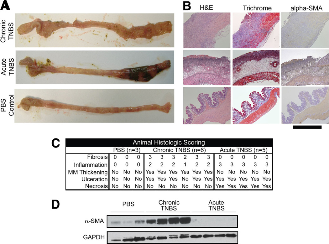
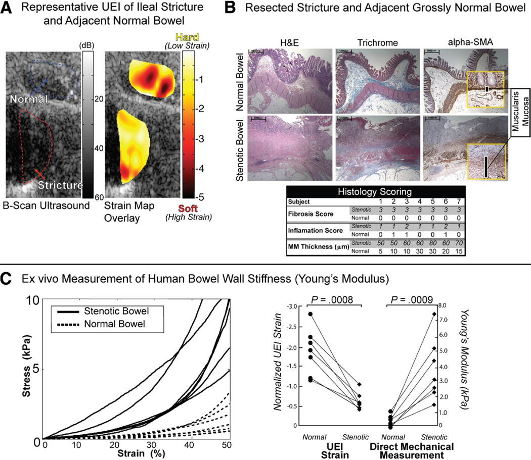
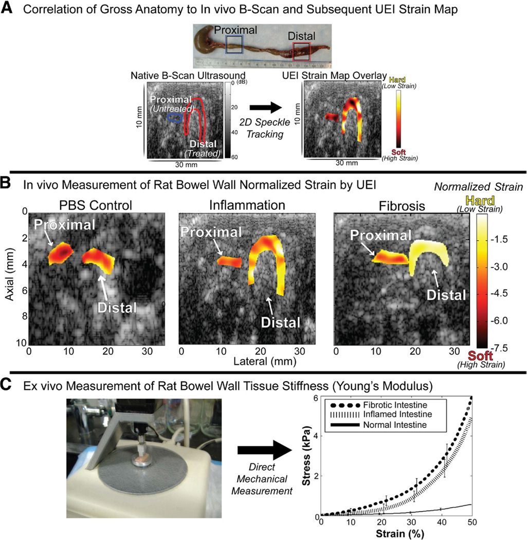
Female Lewis rats underwent weekly trinitrobenzene sulfonic acid enemas yielding models of acute inflammatory colitis (n = 5) and chronic intestinal fibrosis (n = 6). UEI scanning used a novel speckle-tracking algorithm to estimate tissue strain. Resected bowel segments were evaluated for evidence of inflammation and fibrosis. Seven consecutive patients with stenotic CD were studied with UEI and their resected stenotic and normal bowel segments were evaluated by ex vivo elastometry and histopathology.
Transcutaneous UEI normalized strain was able to differentiate acutely inflamed (-2.07) versus chronic fibrotic (-1.10) colon in rat models of inflammatory bowel disease (IBD; P = .037). Transcutaneous UEI normalized strain also differentiated stenotic (-0.87) versus adjacent normal small bowel (-1.99) in human CD (P = .0008), and this measurement also correlated well with ex vivo elastometry (r = -0.81).
UEI can differentiate inflammatory from fibrotic intestine in rat models of IBD and can differentiate between fibrotic and unaffected intestine in a pilot study in humans with CD. UEI represents a novel technology with potential to become a new objective measure of progression of intestinal fibrosis. Prospective clinical studies in CD are needed.
肠道纤维化可导致许多克罗恩病(CD)的并发症。现有的生物标志物和影像学方法缺乏足够的准确性,无法区分肠道炎症和纤维化。经皮超声弹性成像(UEI)是一种有前途的、非侵入性的测量组织力学特性的方法。我们假设 UEI 可以区分结肠炎动物模型和 CD 患者的炎症性和纤维化肠壁变化。
雌性 Lewis 大鼠每周接受三硝基苯磺酸灌肠,产生急性炎症性结肠炎模型(n = 5)和慢性肠道纤维化模型(n = 6)。UEI 扫描使用一种新的斑点跟踪算法来估计组织应变。切除的肠段用于评估炎症和纤维化的证据。对 7 例连续狭窄性 CD 患者进行 UEI 检查,并对其狭窄和正常肠段进行离体弹性测量和组织病理学检查。
经皮 UEI 归一化应变能够区分炎症性结肠炎(IBD)大鼠模型中的急性炎症(-2.07)与慢性纤维化(-1.10)(P =.037)。经皮 UEI 归一化应变也能区分 CD 患者狭窄段(-0.87)与相邻正常小肠(-1.99)(P =.0008),并且该测量与离体弹性测量相关性良好(r = -0.81)。
UEI 可区分 IBD 大鼠模型中的炎症性和纤维化肠道,也可区分 CD 患者纤维化和未受影响的肠道。UEI 代表了一种新的技术,有可能成为肠道纤维化进展的新的客观测量方法。需要在 CD 中进行前瞻性临床研究。