Pain Research Center, Department of Anesthesiology, University of Cincinnati College of Medicine, Cincinnati, OH 45267-0531, USA.
Mol Pain. 2011 Jul 27;7:53. doi: 10.1186/1744-8069-7-53.
Sprouting of sympathetic fibers into sensory ganglia occurs in many preclinical pain models, providing a possible anatomical substrate for sympathetically enhanced pain. However, the functional consequences of this sprouting have been controversial. We used a transgenic mouse in which sympathetic fibers expressed green fluorescent protein, observable in live tissue. Medium and large diameter lumbar sensory neurons with and without nearby sympathetic fibers were recorded in whole ganglion preparations using microelectrodes.
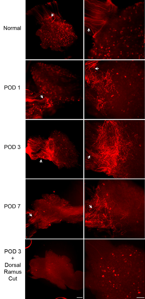
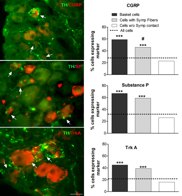
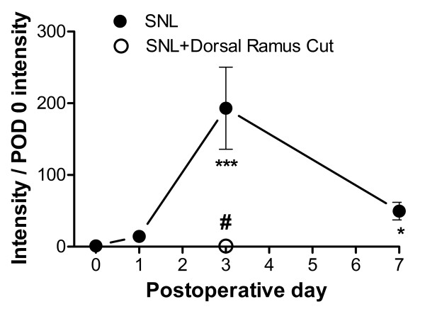
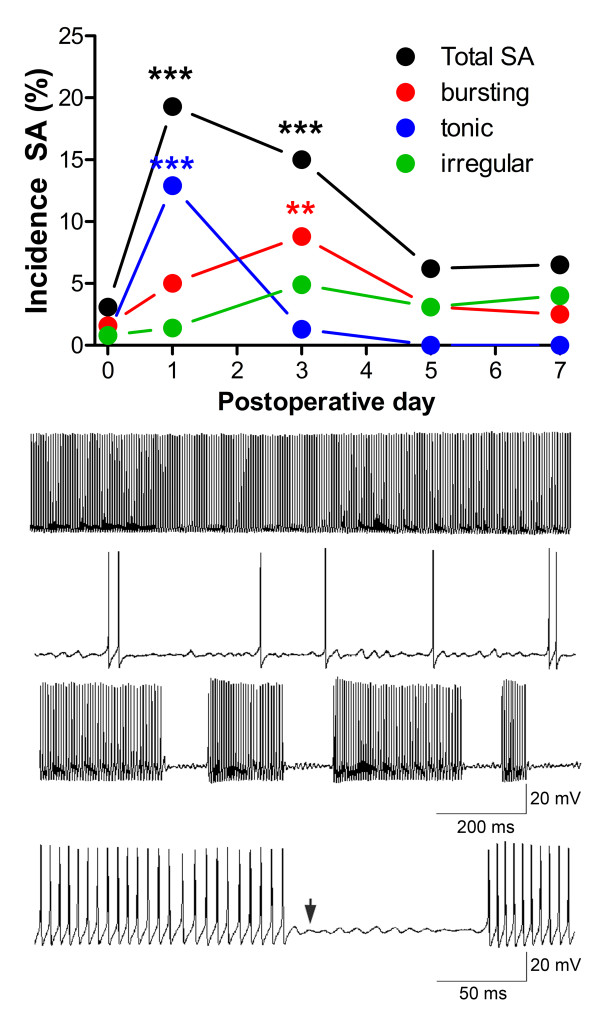
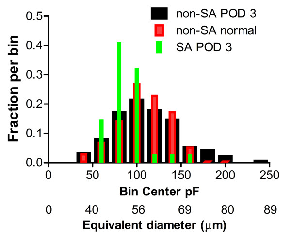
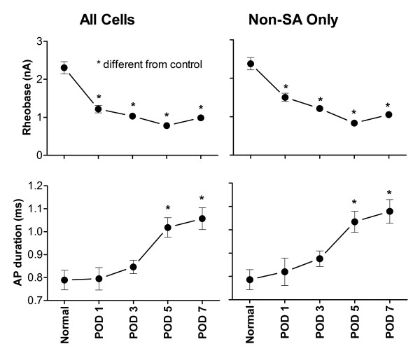
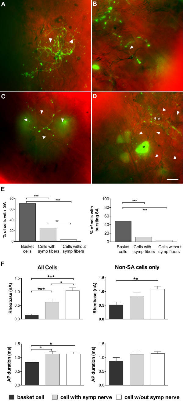
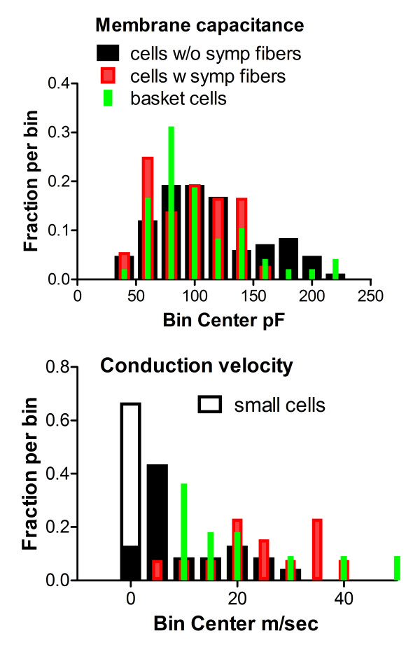
After spinal nerve ligation, sympathetic sprouting was extensive by 3 days. Abnormal spontaneous activity increased to 15% and rheobase was reduced. Spontaneously active cells had Aαβ conduction velocities but were clustered near the medium/large cell boundary. Neurons with sympathetic basket formations had a dramatically higher incidence of spontaneous activity (71%) and had lower rheobase than cells with no sympathetic fibers nearby. Cells with lower density nearby fibers had intermediate phenotypes. Immunohistochemistry of sectioned ganglia showed that cells surrounded by sympathetic fibers were enriched in nociceptive markers TrkA, substance P, or CGRP. Spontaneous activity began before sympathetic sprouting was observed, but blocking sympathetic sprouting on day 3 by cutting the dorsal ramus in addition to the ventral ramus of the spinal nerve greatly reduced abnormal spontaneous activity.
The data suggest that early sympathetic sprouting into the sensory ganglia may have highly localized, excitatory effects. Quantitatively, neurons with sympathetic basket formations may account for more than half of the observed spontaneous activity, despite being relatively rare. Spontaneous activity in sensory neurons and sympathetic sprouting may be mutually re-enforcing.
在许多临床前疼痛模型中,交感神经纤维向感觉神经节发芽,为交感神经增强疼痛提供了可能的解剖学基础。然而,这种发芽的功能后果一直存在争议。我们使用了一种转基因小鼠,其中交感神经纤维表达绿色荧光蛋白,可在活体组织中观察到。使用微电极在整个神经节制剂中记录有和没有附近交感神经纤维的中大和大直径腰椎感觉神经元。
在脊神经结扎后,3 天内交感神经发芽广泛。异常自发性活动增加到 15%,并且阈值降低。自发性活动细胞具有 Aαβ传导速度,但聚集在中/大细胞边界附近。具有交感神经篮状结构的神经元自发性活动发生率显著升高(71%),并且比附近没有交感神经纤维的细胞阈值更低。附近纤维密度较低的细胞具有中间表型。对切片神经节的免疫组织化学染色显示,被交感神经纤维包围的细胞富含伤害性标记物 TrkA、P 物质或 CGRP。自发性活动在观察到交感神经发芽之前就开始了,但是在第 3 天通过切断脊神经的背支除了腹支来阻断交感神经发芽,可以大大减少异常自发性活动。
这些数据表明,早期交感神经向感觉神经节的发芽可能具有高度局部的兴奋作用。从数量上看,尽管相对较少,但具有交感神经篮状结构的神经元可能占观察到的自发性活动的一半以上。感觉神经元的自发性活动和交感神经发芽可能相互促进。