Young K D, Erickson K, Nugent A C, Fromm S J, Mallinger A G, Furey M L, Drevets W C
National Institutes of Health, National Institute of Mental Health, Section on Neuroimaging in Mood and Anxiety Disorders, Bethesda, MD, USA.
Psychol Med. 2012 Feb;42(2):345-57. doi: 10.1017/S0033291711001371. Epub 2011 Jul 29.
Major depressive disorder (MDD) is associated with deficits in recalling specific autobiographical memories (AMs). Extensive research has examined the functional anatomical correlates of AM in healthy humans, but no studies have examined the neurophysiological underpinnings of AM deficits in MDD. The goal of the present study was to examine the differences in the hemodynamic response between patients with MDD and controls while they engage in AM recall.
Participants (12 unmedicated MDD patients; 14 controls) underwent functional magnetic resonance imaging (fMRI) scanning while recalling AMs in response to positive, negative and neutral cue words. The hemodynamic response during memory recall versus performing subtraction problems was compared between MDD patients and controls. Additionally, a parametric linear analysis examined which regions correlated with increasing arousal ratings.
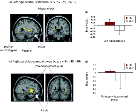
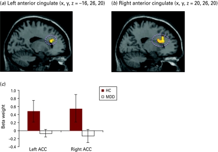
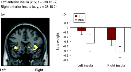
Behavioral results showed that relative to controls, the patients with MDD had fewer specific (p=0.013), positive (p=0.030), highly arousing (p=0.036) and recent (p=0.020) AMs, and more categorical (p<0.001) AMs. The blood oxygen level-dependent (BOLD) response in the parahippocampus and hippocampus was higher for memory recall versus subtraction in controls and lower in those with MDD. Activity in the anterior insula was lower for specific AM recall versus subtraction, with the magnitude of the decrement greater in MDD patients. Activity in the anterior cingulate cortex was positively correlated with arousal ratings in controls but not in patients with MDD.
We replicated previous findings of fewer specific and more categorical AMs in patients with MDD versus controls. We found differential activity in medial temporal and prefrontal lobe structures involved in AM retrieval between MDD patients and controls as they engaged in AM recall. These neurophysiological deficits may underlie AM recall impairments seen in MDD.
重度抑郁症(MDD)与特定自传体记忆(AMs)回忆缺陷有关。大量研究已探讨了健康人自传体记忆的功能解剖学相关性,但尚无研究考察MDD患者自传体记忆缺陷的神经生理学基础。本研究的目的是在MDD患者和对照者进行自传体记忆回忆时,检查他们血液动力学反应的差异。
参与者(12名未服药的MDD患者;14名对照者)在根据积极、消极和中性提示词回忆自传体记忆时接受功能磁共振成像(fMRI)扫描。比较了MDD患者和对照者在记忆回忆与进行减法运算时的血液动力学反应。此外,进行了参数线性分析,以检查哪些区域与唤醒评分增加相关。
行为结果显示,与对照者相比,MDD患者的特定自传体记忆(p = 0.013)、积极自传体记忆(p = 0.030)、高唤醒自传体记忆(p = 0.036)和近期自传体记忆(p = 0.020)较少,而类别性自传体记忆较多(p < 0.001)。对照者在海马旁回和海马中,记忆回忆时的血氧水平依赖(BOLD)反应高于减法运算时,而MDD患者则较低。在特定自传体记忆回忆与减法运算时,前岛叶的活动较低,MDD患者中这种下降幅度更大。前扣带回皮层的活动在对照者中与唤醒评分呈正相关,而在MDD患者中则不然。
我们重复了之前的研究结果,即MDD患者与对照者相比,特定自传体记忆较少,类别性自传体记忆较多。我们发现,MDD患者和对照者在进行自传体记忆回忆时,参与自传体记忆提取的内侧颞叶和前额叶结构存在不同的活动。这些神经生理学缺陷可能是MDD患者自传体记忆回忆受损的基础。