Jana Manisha, Srivastava Deep Narayan, Sharma Raju, Gamanagatti Shivanand, Nag Hiralal, Mittal Ravi, Upadhyay Ashish Dutt
Department of Radiodiagnosis, All India Institute of Medical Sciences, Ansari Nagar, New Delhi, India.
Indian J Radiol Imaging. 2011 Apr;21(2):98-106. doi: 10.4103/0971-3026.82284.
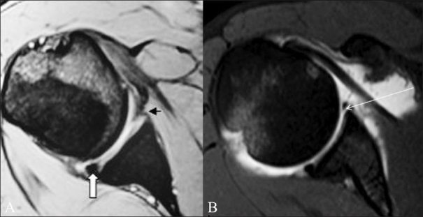
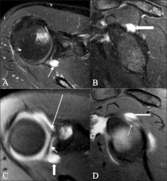
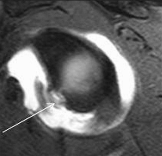
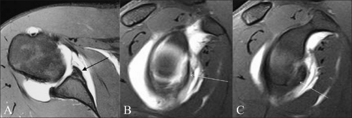
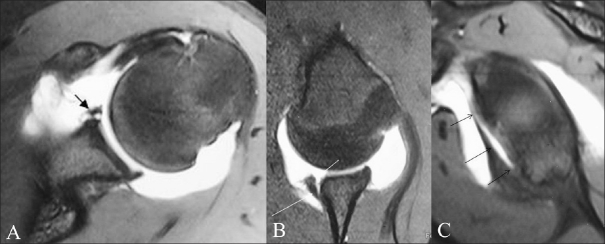
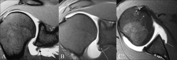
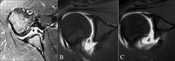
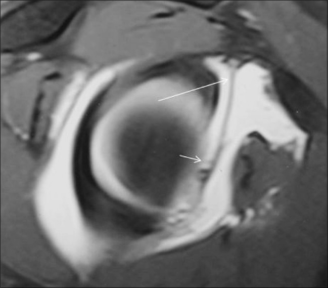
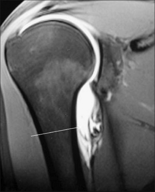
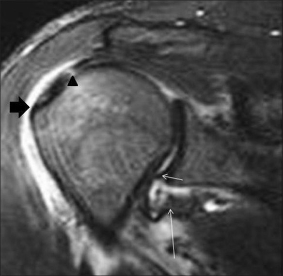
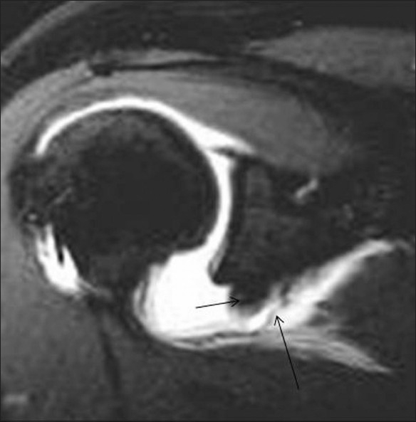
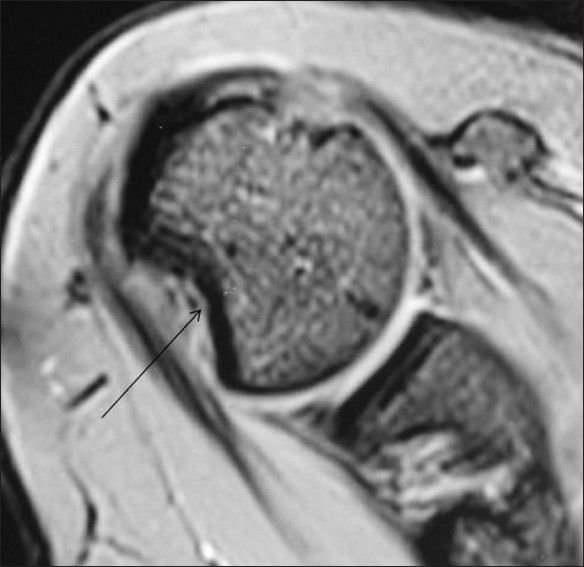
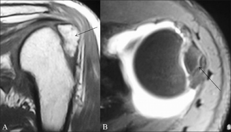
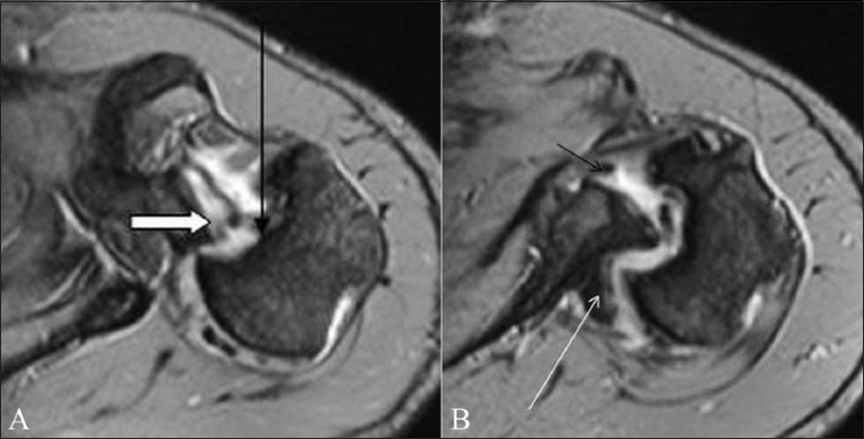
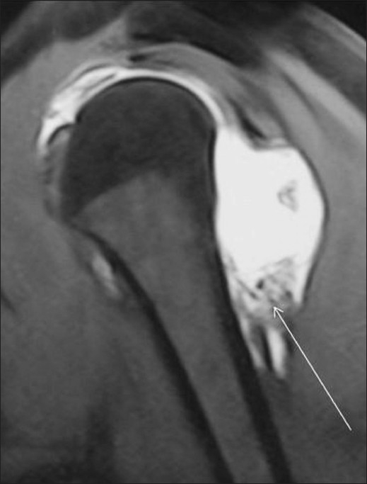
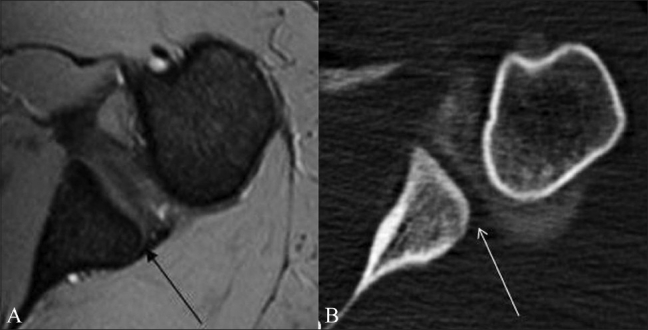
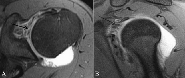
The glenohumeral joint is the most commonly dislocated joint in the body, and anterior instability is the most common type of shoulder instability. Depending on the etiology and the age of the patient, there may be associated injuries, for example, to the anterior-inferior labro-ligamentous structures (in young individuals with traumatic instability) or to the bony components (commoner in the elderly), which are best visualized using MRI and MR arthrography. Anterior instability is associated with a Bankart lesion and its variants and abnormalities of the anterior band of the inferior glenohumeral ligament (IGHL), whereas posterior instability is associated with reverse Bankart and reverse Hill-Sachs lesions. Cases of multidirectional instability often have no labral pathology on imaging but show specific osseous changes including increased chondrolabral retroversion. This article reviews the relevant anatomy in brief and describes the MRI findings in each type, with the imaging features of the common abnormalities.
盂肱关节是人体最常发生脱位的关节,而前向不稳定是肩部不稳定最常见的类型。根据病因和患者年龄,可能会伴有其他损伤,例如,前下盂唇韧带结构损伤(见于有创伤性不稳定的年轻个体)或骨质成分损伤(在老年人中更常见),这些损伤最好通过磁共振成像(MRI)和磁共振关节造影来观察。前向不稳定与Bankart损伤及其变异以及下盂肱韧带(IGHL)前束异常有关,而后向不稳定与反Bankart和反Hill-Sachs损伤有关。多向不稳定病例在影像学上通常没有盂唇病变,但会显示特定的骨质改变,包括软骨盂唇后倾增加。本文简要回顾相关解剖结构,并描述每种类型的MRI表现以及常见异常的影像学特征。