Laboratório de Citogenética Humana, Instituto de Ciências Biológicas, Universidade Federal do Pará, Belém, Brazil.
PLoS One. 2011;6(7):e21988. doi: 10.1371/journal.pone.0021988. Epub 2011 Jul 21.
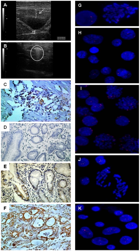
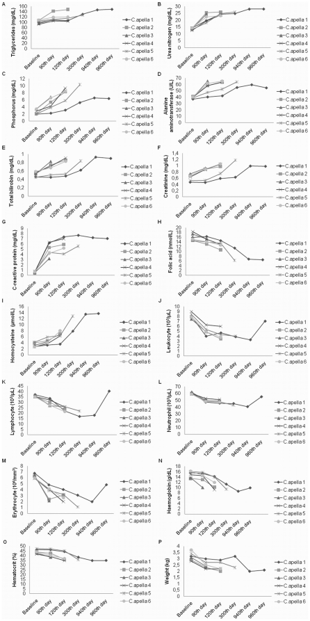
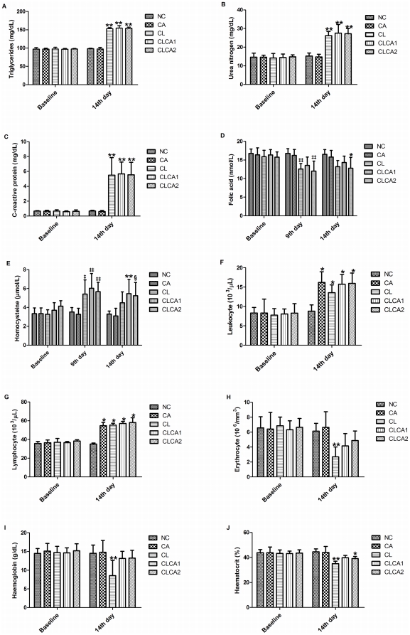
The evolution of gastric carcinogenesis remains largely unknown. We established two gastric carcinogenesis models in New-World nonhuman primates. In the first model, ACP03 gastric cancer cell line was inoculated in 18 animals. In the second model, we treated 6 animals with N-methyl-nitrosourea (MNU). Animals with gastric cancer were also treated with Canova immunomodulator. Clinical, hematologic, and biochemical, including C-reactive protein, folic acid, and homocysteine, analyses were performed in this study. MYC expression and copy number was also evaluated. We observed that all animals inoculated with ACP03 developed gastric cancer on the 9(th) day though on the 14(th) day presented total tumor remission. In the second model, all animals developed pre-neoplastic lesions and five died of drug intoxication before the development of cancer. The last surviving MNU-treated animal developed intestinal-type gastric adenocarcinoma observed by endoscopy on the 940(th) day. The level of C-reactive protein level and homocysteine concentration increased while the level of folic acid decreased with the presence of tumors in ACP03-inoculated animals and MNU treatment. ACP03 inoculation also led to anemia and leukocytosis. The hematologic and biochemical results corroborate those observed in patients with gastric cancer, supporting that our in vivo models are potentially useful to study this neoplasia. In cell line inoculated animals, we detected MYC immunoreactivity, mRNA overexpression, and amplification, as previously observed in vitro. In MNU-treated animals, mRNA expression and MYC copy number increased during the sequential steps of intestinal-type gastric carcinogenesis and immunoreactivity was only observed in intestinal metaplasia and gastric cancer. Thus, MYC deregulation supports the gastric carcinogenesis process. Canova immunomodulator restored several hematologic measurements and therefore, can be applied during/after chemotherapy to increase the tolerability and duration of anticancer treatments.
胃癌的发生机制在很大程度上尚不清楚。我们在新世界非人灵长类动物中建立了两种胃癌发生模型。在第一种模型中,将 ACP03 胃癌细胞系接种于 18 只动物。在第二种模型中,我们用 N-甲基-N-亚硝脲(MNU)处理 6 只动物。患有胃癌的动物也接受 Canova 免疫调节剂治疗。本研究进行了临床、血液学和生化分析,包括 C 反应蛋白、叶酸和同型半胱氨酸。还评估了 MYC 表达和拷贝数。我们观察到,所有接种 ACP03 的动物在第 9 天均发生胃癌,但在第 14 天全部肿瘤完全消退。在第二种模型中,所有动物均发生癌前病变,5 只动物因药物中毒在癌症发生前死亡。最后一只幸存的 MNU 处理动物在第 940 天通过内窥镜检查发现肠型胃腺癌。在 ACP03 接种动物和 MNU 处理中,随着肿瘤的出现,C 反应蛋白水平和同型半胱氨酸浓度增加,而叶酸水平降低。ACP03 接种还导致贫血和白细胞增多。血液学和生化结果与胃癌患者观察到的结果相符,支持我们的体内模型可能有助于研究这种肿瘤。在细胞系接种动物中,我们检测到 MYC 免疫反应性、mRNA 过表达和扩增,这与体外观察结果一致。在 MNU 处理动物中,在肠型胃癌发生的连续步骤中,mRNA 表达和 MYC 拷贝数增加,而免疫反应性仅在肠上皮化生和胃癌中观察到。因此,MYC 失调支持胃癌的发生过程。Canova 免疫调节剂恢复了多项血液学测量值,因此可在化疗期间/之后应用,以提高抗癌治疗的耐受性和持续时间。